INTRODUCTION
Skin avulsions are severe traumatic injuries, in which sections of skin and subcutaneous tissue are torn off from the body. Once an avulsed flap is detached from the body, the microcirculation of subdermal capillaries is immediately impaired and skin necrosis of the avulsed area will happen [1]. The surgical management and salvage of avulsion injuries are quite challenging for surgeons due to their high morbidity and mortality [2]. When the loss of an avulsed flap due to necrosis is too extensive, secondary operative procedures may be required. Compared to complete salvage of an avulsed flap through primary repair, secondary-intention wound healing or skin coverage via a secondary operation is accompanied by a severe postoperative scar. In this case, a severe avulsion injury of the nose happened due to a traumatic accident. To minimize the loss of the distal portion of the avulsed flap, we used a combination of three treatments: polydeoxyribonucleotide (PDRN) injection, continuous non-rebreather mask oxygen therapy, and chemical leeching. We report an excellent outcome of using this triple-combination treatment for maximizing salvage of an avulsed nasal chondrocutaneous flap.
CASE REPORT
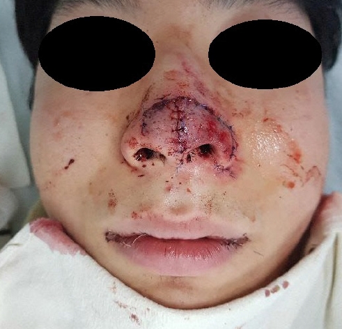
A 24-year-old male patient presented to the emergency room with a severe traumatic avulsion injury on his nose. The patient was a non-smoker and did not have any comorbidities. The patient fainted right after a heavy deadlift and fell forward onto the barbell. His nose grazed the rough surface of the barbell and an avulsion injury took place. The avulsion injury caused two avulsed flaps (Fig. 1A). The right avulsed flap was smaller, and only the subcutaneous layer was involved (Fig. 1B). The left avulsed flap was larger and involved all layers of the middle to lower nose, including the nasal skin, lower lateral cartilage, and nasal mucosa (Fig. 1C). Fortunately, the columellar artery was not damaged despite a longitudinal laceration on the columella. The overall flap color was already purplish upon the patient’s initial presentation.
Primary repair of the nasal cartilage and subcutaneous tissue was done with Vicryl 6-0 (Ethicon). Skin repair was done with Prolene 6-0 (Ethicon) in the emergency room under topical anesthesia, and microvascular anastomosis was not performed (Fig. 2). Only a key suture was done in the nasal mucosa, and it was left semi-open for 2 days to let out subsequent bleeding. To maintain the alignment of nasal cavity, Merocel (Medtronic Xomed Inc.) packing was done and changed once a day. Two days after the primary repair, mucosal bleeding subsided, so we repaired the mucosal dehiscence completely.
Next, half a vial of Placentex (Pharma Research Co., Ltd.; PDRN sodium 5.625 mg) was evenly injected on the subcutaneous layer of both the damaged and undamaged sides of the nose using a 26-gauge needle once a day for 3 days. The injected volume at each point was minimal to avoid making a fluid collection under the avulsed flap. One vial of Placentex was also systemically administered intramuscularly twice a day for 7 days.
Continuous non-rebreather mask oxygen therapy was applied on the patient’s face tightly immediately after the primary repair. Humidified oxygen was supplied to prevent the flap from drying. The oxygen flow rate was kept at 15 L/min continuously for 7 days. Except during wound dressing or eating meals, the patient was asked not to take off the mask.
To manage the acute venous congestion of the flap, we also used the chemical leeching technique, for which 0.5 vial of heparin sodium (5,000 IU; Choongwae) was mixed with 5 mL of normal saline. A subdermal injection of this diluted heparin was performed at on several points of the distal portion of the avulsed flap using a 26-gauge needle immediately after the primary repair. After the injections on the flap, pin-point bleeding from the injected site was observed. Furthermore, the blood clots along the wound margin were not solidified due to the antithrombotic effect of heparin and could be easily removed by gentle rinsing or rubbing. After cleansing the blood clots on the wound margin, the diluted heparin solution was spread evenly on the flap and along the wound margin. Effexin ointment (Ildong) containing ofloxacin was then applied to the flap. Next, the wound was covered with Vaseline gauze for protection and moisturizing. This dressing procedure was performed three times a day for the first 3 days and two times a day on postoperative days 4 and 5. Intravenous heparin was not administered.
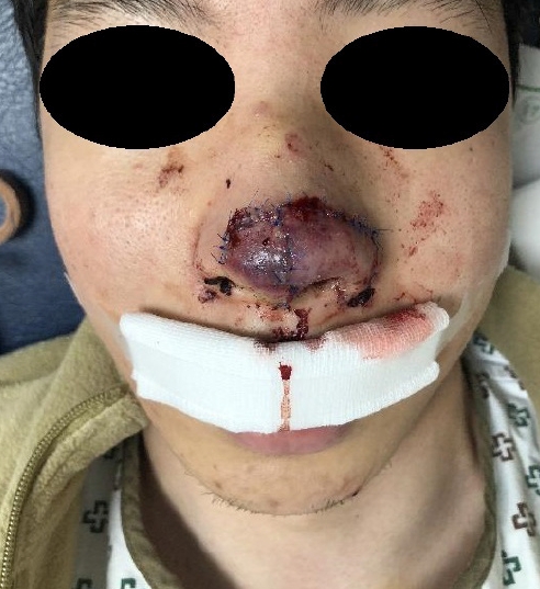
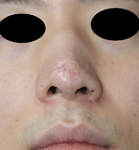
For the first 2 days after the primary repair, venous congestion of the avulsed flap seemed to be progressing (Fig. 3). The amount of bleeding from the wound started to decrease on postoperative day 3 and disappeared on postoperative day 5. The distal flap color gradually changed from dark purple to bright red (Fig. 4) and slowly recovered to the normal skin color (Fig. 5). Overall, the avulsion wound fully recovered without any major deformity or functional impairment of the nose (Fig. 6). The patient showed full satisfaction in his last visit on postoperative day 540.
DISCUSSION
As the nose is the most prominent feature of the face, it is prone to be injured by external damage. Among various types of traumatic injuries of the nose, an avulsion injury can cause an incidental nasal chondrocutaneous flap because the nose is made of a cartilaginous framework and skin envelope. When an avulsion injury happens on the nose, the subdermal capillaries of the avulsed flap are detached and the microcirculation of the skin and subcutaneous tissue is disrupted [1]. Although there is intact arterial perfusion for the flap, venous congestion of the distal region of the flap usually happens because of the disrupted microcirculation along the edge. Subsequent small-vessel venous thrombosis occurs, leading to distal flap loss due to venous insufficiency, which leads to necrosis [3]. As neovascularization at the wound margin happens when the endothelial cells are able to proliferate and migrate to the wound [4], a solid hematoma mechanically disturbing the close connection of the dermis layer can interrupt the neovascularization of the edge. Moreover, the cartilage of the nasal chondrocutaneous flap acts as a mechanical barrier that limits the perpendicular vascularity of the flap [5]. Thus, an accurate dermis connection to the wound margin dermis for horizontal neovascularization is particularly important for the viability of a nasal chondrocutaneous flap [5].
To maintain an accurate dermis connection along the wound margin, adequate primary repair should be performed first. After primary repair, hematoma removal and prevention of solid hematoma formation inside the wound margin are very important. Therefore, we did not completely repair the mucosal layer; instead, only a key suture was done at first to let the bleeding flow out. Two days after the primary repair, intranasal bleeding subsided and we completely repaired the mucosal layer.
Medicinal leeches have been used to resolve venous congestion of flaps [6]. However, leeches are tricky to control, and infectious complications of leeches have been reported [7]. Therefore, we used local subcutaneous heparin injection with scarification (i.e., chemical leeching) instead and adopted the protocol using low-molecular-weight heparin from the study of Perez et al. [8]. Fortunately, the venous congestion resolved well. We did not use systemic heparinization because Barnett et al. [9] reported that it may be harmful to patients.
Baynosa and Zamboni [10] reported that hyperbaric oxygen therapy combined with leeching resulted in a significant improvement in flap survival compared to using leeching alone. However, hyperbaric oxygen therapy is expensive, cannot be applied continuously, and poses a risk of oxygen toxicity [11]. Chan and Campbell [11] reported the usability of topical oxygen therapy as an alternative modality for promoting wound healing. We used a nonrebreather mask as a normobaric mask supplying pressurized oxygen to the nose. Applying oxygen at 15 L/min via a non-rebreather mask works as a topical oxygen chamber, increasing the fraction of inspired oxygen (FiO2) from 21% to 70% [12]. According to Gadrey et al. [13], the partial pressure of arterial oxygen is proportionate to FiO2 when no positive pressure ventilation is applied to a patient at room temperature. Thus, increasing the FiO2 by using a non-rebreather mask will supply additional oxygen systemically. By using this modality, we could continuously oxygenate the damaged hypoxic tissue by both diffusion and a systemic supply.
Recent studies have reported that PDRN can accelerate angiogenesis and increase the vessel density of the wound margin. Many previous studies have shown that PDRN can promote cell migration and growth, induce neovascularization, and reduce inflammation in impaired wounds, and its mechanism has been proposed to involve activation of the adenosine receptor A2A and upregulation of vascular endothelial growth factor [14]. In a comparative study using rats, Polito et al. [15] found that ischemic flaps treated with PDRN showed complete recovery, with remarkable increases in blood flow compared to untreated flaps based on quantitative observations using laser Doppler images. Using local and systemic PDRN administration accelerated angiogenesis and wound healing in the avulsed wound margin.
Considering the reasons described above, complete salvage of an avulsed flap without any loss of the distal margin of the flap is extremely difficult. When entire or partial flap loss happens, the resulting scar can be conspicuous, and additional surgery, such as skin grafts, local flaps, or even composite grafts, might be required. Additional costs and recovery time may be imposed on patients as well. Donor site morbidities from a secondary operation can be another problem. Thus, maximizing the salvage of avulsion injuries is quite important.
In this case, the avulsion injury was so severe that the cutting damage involved the entire layer of the patient’s left middle to lower nose. If adequate treatments were not provided at the right time, complete salvage of the avulsed flap might not have been achieved. As shown by the excellent outcome of this case, using three treatments in combination seems to have been effective for minimizing the necrotizing loss of an avulsed flap. However, our study is confined to just one case of a severe avulsion injury. Thus, a large-scale study for statistical verification of the usefulness of this combined treatment strategy for avulsion injuries would be necessary in the future.