INTRODUCTION
Cases of damage to the auricle after being bitten by a person are rare. If the damaged part is not as large as the size of the perpetrator’s mouth, the operator may consider performing an auricular composite graft. However, auricular reconstruction can be challenging because the cartilage and soft tissues are often crushed, and insufficient blood supply in the crushed tissues renders them susceptible to infection. Herein, we report a case in which a human-bitten auricle was reconstructed using two-stage cartilage preservation surgery.
CASE REPORT
A 50-year-old male patient was brought to our emergency room after being bitten by his girlfriend on his right ear. One-third of his ear auricle was completely severed. His girlfriend carried the severed part of the auricle to the emergency room. The amputated part was cleaned with saline, and emergency composite grafting was performed. On his next visit to the plastic surgery clinic, the sutured part appeared black and necrotic (Fig. 1). Therefore, we suspected a composite graft failure. Since the patient experienced intermittent pain and edema in the right auricle, debridement of the necrotic tissue and auricular reconstruction were suggested.
The amputated part spanned the helix and scapha and included some cartilage (3×2 cm). After appropriate induction of general anesthesia, the amputated auricle was removed (Fig. 2). Although the blackish skin was necrotized, the cartilage was not necrotized. The amputated part was sufficiently washed with normal saline, and we removed the skin of the amputee using Metzenbaum scissors to preserve the perichondrium of the cartilage. The cartilage in the amputated part was pulverized from the bite.
The right auricle was pushed toward the scalp to mark the incision line to help bury the skinned cartilage. We incised the postauricular skin at the contact point where the skinned cartilage met the postauricular skin. After dissecting the subcutaneous plane to create a pocket of appropriate size, the de-epithelialized cartilage was carefully inserted under the postauricular skin (Fig. 3). The incision line was sutured to the skin of the right ear. To ensure proper contact between the right ear and postauricular skin, a mild compression dressing combined with an ointment and gauze was used. The dressing was placed to maintain the position of the parts and immobilize the suture site. The dressing was precisely fitted and supported using tape strips.
Separation surgery was performed on the 14th day after the burying surgery. The incision line was marked to cover the buried cartilage. The postauricular skin and cartilage connected to the right ear were elevated in one block (Fig. 4). The postauricular skin donor site was covered with a split-thickness skin graft from the thigh.
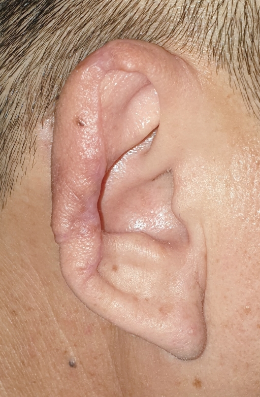
The patient was discharged one day after surgery and received outpatient treatment, which included oral antibiotic treatment (Mexesin capsules; 500 mg; three times per day) and dressing changes on alternate days in the outpatient clinic. The wound was healing well 1 week after the operation. There were no signs of hyperemia or infection. No sulcus formation was visualized at that time. The suture was removed 2 weeks after surgery, and there were no signs of necrosis or exposed cartilage. A sulcus had started forming between the helix and triangular fossa. The wound had completely healed 1 month after surgery and had an excellent contour helix (Fig. 5).
There was no hypertrophic scar at 3 months postoperatively. We observed a normal ear contour that harmonized well with the surrounding skin color.
DISCUSSION
Reports of human bite injuries that cause auricular damage are relatively rare in the literature. Auricular reconstruction can be challenging. Microvascular anastomosis is ideal in cases of complete auricular amputation. However, Kind et al. [1] reported that the small inner diameter of the vessels in the outer ear makes anastomosis difficult. Moreover, microvascular anastomosis prolongs the operation time, and venous anastomosis is often impossible in practice [2]. Even after successful anastomosis, further salvage therapy using medical leeches or venous blood loss to ease venous congestion may be required. Microvascular anastomosis causes inconvenience to the patient or medical staff and lengthens the treatment period.
In contrast, a composite graft (simple sutures without microanastomosis) is a relatively simple method. The major disadvantage of composite grafts is graft failure, especially when the contact surface of the amputated segment is small [3].
Creating stable contact of the cut end with the surrounding tissues over a large area through re-epithelialization can help ensure a sufficient supply of blood and serum, thereby increasing the survival rate of grafts [4]. In addition, unlike microvascular anastomosis, composite grafting allows the patient to be discharged within a few days without the need for special surveillance or prescription of anticoagulants, since there is no risk of venous congestion. Moreover, because the auricular cartilage and dermis are preserved, there is no deformation due to the absence of the cartilage skeleton.
After the wound is healed, epithelium of the same color is produced naturally in good harmony with the surrounding skin, and wound management is convenient. Rapid recovery of blood circulation prevents the formation of a large area of scar tissue. This can further minimize the deformation of the auricular cartilage caused by scar formation.
Despite the excellent outcomes, several aspects must be considered when treating a case of auricular amputation with a scalp flap. The flap size must be adequate to cover the defect, and positioning the remnant ear auricle on the scalp is sometimes challenging. Inappropriate positioning can lead to flap detachment from the remnant ear auricle. Therefore, proper contact between the ear skin tip and the postauricular skin is crucial.
If the perpetrator is closely related to the patient, the perpetrator may feel more anxious and sensitive about the treatment process than the patient and may not be satisfied with the postoperative outcome. In our case, the perpetrator was the patient’s girlfriend, and she was mildly dissatisfied with the surgical outcome. She wanted a perfect surgical result in order to overcome her guilt. Medical staff must help the perpetrator and patient re-establish their relationship, and should take care not to worsen the relationship between the perpetrator and patient.
Using two-stage cartilage preservation surgery, we successfully reconstructed a severely gashed auricle and achieved cosmetically and functionally satisfactory results.
In conclusion, after the failure of a composite graft in a patient with complete auricle amputation, we performed two-stage cartilage preservation surgery and successfully reconstructed the severely gashed auricle and achieved cosmetically and functionally satisfactory results.