Rabbit model for the study of nasal tip plasty: a comparative cephalometric analysis after rhinoplasty
Article information

Abstract
Background
Open rhinoplasty and septoplasty have emerged as popular surgical techniques for both functional and aesthetic procedures. To study open rhinoplasty with or without septoplasty, the use of animals is necessary. However, no reports have been published on radiologic methods for evaluating rhinoplasty or nasal tip plasty in animals using cephalometry. In this study, a validated model of open rhinoplasty and septoplasty was utilized in rabbits to establish radiographic guidelines for assessing the degree of tip plasty in these animals.
Methods
Eight adult New Zealand rabbits were used to establish an autologous septal extension graft (SEG) model. The rabbits underwent concurrent submucosal septal resection and open rhinoplasty. The SEG was implanted using nasal septal cartilage. To evaluate the results of nasal tip plasty, lateral-view X-ray images were obtained preoperatively, as well as 2 days, 2 months, and 12 months postoperatively.
Results
This open rhinoplasty rabbit model appears to be a practical tool for studying open rhinoplasty and tip plasty, demonstrating statistically significant results following SEG implantation. Furthermore, it is suitable for training purposes, specifically for the submucosal resection of septal cartilage.
Conclusions
This study presents a statistical analysis of the long-term (1-year) postoperative results of SEG implantation, using experimental procedures like those utilized in humans. Through a cephalometric comparison of rabbit noses, the effect of various SEG and tip plasty methods on the rabbit nose can be objectively measured.
INTRODUCTION
Rhinoplasty can be categorized into two types: open and closed. Open rhinoplasty, with or without septoplasty, has emerged as a highly favored procedure for both aesthetic and functional reasons. Numerous complex techniques for open rhinoplasty have been suggested, but comparing results across humans presents a challenge [1]. Consequently, a need exists for animal study models to compare objective results. Rabbits, which possess a suitable amount of septal cartilage, are routinely used in animal experiments as the primary animal model for investigating nasal surgery [2]. This study introduces animal models for open rhinoplasty and autologous septoplasty that allow access to the nasal columella and septum for septal extension graft (SEG) implantation.
The radiologic evaluation of a rhinoplasty model is valuable, primarily because the rabbit nose, unlike the human nose, is convex (Fig. 1) [3]. This characteristic makes it impossible to adequately assess the nasal dorsum based solely on external appearance. The conventional method involves shaving the hair from the rabbit’s snout and then evaluating the results through photography. However, this process of hair removal could introduce variations in the results obtained. Consequently, we suggest employing a cephalometric analysis using X-ray images. This approach allows for an objective evaluation, irrespective of the presence of hair or the shape of the snout.

(A) The first reference line was established by drawing a line from the nasion to the premolar teeth. The second reference line was drawn from the nasion to the defining point of the tip. Digital measurements were then taken of the angle formed between the first and second reference lines. (B) Human cephalometric model.
METHODS
In this prospective animal study, eight 6- to 8-month-old New Zealand white rabbits, each weighing between 3.5 and 4 kg, were used. All rabbits that underwent surgery were administered 50 mg/kg of ketamine hydrochloride and 5 mg/kg of xylazine for general anesthesia [4]. Following sedation and orotracheal intubation, local anesthesia, which consisted of lidocaine and 2% epinephrine hydrochloride (1:100,000), was injected. The mucoperichondrium of the septum and columella were infiltrated. A reverse V-shaped transcolumellar incision was made and subsequently connected to the marginal incisions (Fig. 2). The columellar flap and the bilateral mucoperichondrium of the septum were elevated, leaving behind a 0.4-cm L-shaped segment of caudal and dorsal cartilage to support the lower part of the nose. This segment of the nasal septal cartilage was then harvested (Fig. 3). After the septal cartilage was collected, the SEG was positioned perpendicular to the nasal dorsum (Fig. 4). In this study, we did not use a dorsal nasal implant. Notably, however, a dorsal implant made from a material such as silicone can be easily utilized. After securing the nasal septal cartilage on the remaining L strut, the columellar flap was repaired. For analysis of the results, lateral-view X-ray images were obtained preoperatively and at 2 days, 2 months, and 12 months postoperatively. For the cephalometric analysis, a line was drawn from the nasion to the medial line of the first premolar teeth as the first reference line. The second reference line was drawn from the nasion to the defining point of the tip [5,6]. Digital measurements of the angle between the first and second reference lines were then obtained (Fig. 4).

Intraoperative view illustrating the dissection plane after the creation of the transcolumellar and marginal incisions.
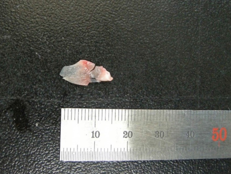
Rabbit septal cartilage of approximately 1.5×0.7 cm. Septal cartilage was used for the septal extension graft.

Intraoperative view of the autogenous bone strut graft. The graft was fixed with Vicryl 5-0 through a hole in the bone.
Statistical analyses were performed using SPSS version 23.0 (IBM Corp.), with P-values of less than 0.05 considered to indicate statistical significance. The Wilcoxon signed-rank test was employed to assess the data.
RESULTS
Eight rabbits were used in this study. No rabbits died, and all tolerated surgery and exhibited normal growth. The Wilcoxon signed-rank test, along with the corresponding P-values, demonstrated a statistically significant increase in the angle between the first and second reference lines (Fig. 5) following open rhinoplasty using an SEG (Fig. 6).

Preoperative and postoperative results in rabbits. Values are presented as mean with standard deviation.

After septal extension graft implantation with septal cartilage. The first reference line remains largely unchanged from before the operation; however, the second reference line has shifted anteriorly, increasing the angle between the first and second reference lines. (A) Preoperative cephalometric analysis of a lateral-view X-ray image of rabbit 1 and (B) cephalometric analysis of a lateral-view X-ray image taken 12 months postoperatively. (C) Preoperative cephalometric analysis of a lateral-view X-ray image of rabbit 2 and (D) cephalometric analysis of a lateral-view X-ray image taken 12 months postoperatively.
A statistically significant difference was observed between the preoperative results and those obtained 2 days postoperatively (P < 0.05). However, no such difference was noted between the 2-day and 12-month postoperative results (P > 0.05). This indicates that the overall outcomes were comparable at 2 days and 12 months following the surgical procedure (Table 1).
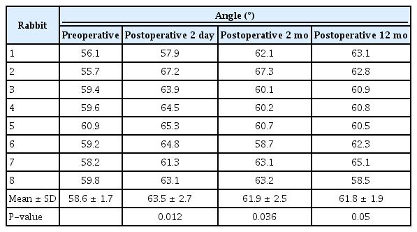
Raw data of preoperative and postoperative results in rabbits
DISCUSSION
Rhinoplasty is among the most frequently performed procedures in the field of plastic surgery. However, postoperative complications such as hemorrhage, hematoma, infection, scarring, patient dissatisfaction, and the need for revision surgery continue to pose challenges for plastic surgeons [1,3,7]. Occasionally, grafts are required for augmentation in both primary and secondary cases, serving both functional and aesthetic purposes [1]. A functional application of this is the use of an SEG at the internal nasal valve during rhinoplasty [8]. Aesthetic applications include nasal lengthening, tip projection, and rotation [1]. The septal cartilage is a commonly used resource for SEG in short nose correction. It is widely employed as the donor material because it directly extends and provides robust support to the alar cartilage. Furthermore, it is readily available in the same surgical field. The SEG is an effective method for tip projection and lengthening in rhinoplasty, first described by Byrd et al. [9]. Subsequently, alternative methods have been evaluated, including the tongue-and-groove technique proposed by Guyuron and Varghai [10,11]. For East Asian people, the SEG has become the preferred technique for augmentation rhinoplasty, due to its superior capacity to correct tip projection and rotation [12].
Despite numerous studies published on the long-term outcomes of grafts used in nasal procedures, ongoing debate remains about the optimal material to employ. This disagreement seems to stem largely from the challenges associated with thoroughly assessing the materials once they have been implanted. The selection of the most suitable model is of paramount importance for standardizing the procedure [3].
Our findings indicate that the implantation of an SEG significantly alters the shape of the nasal tip compared to its preoperative state, thereby demonstrating the effectiveness of the SEG technique. The results further illustrate the durability of the nasal SEG, even 12 months postoperation. Consequently, we can infer that an SEG can induce a long-term modification in the shape of the nasal tip. It is also evident that nasal septal surgery and open rhinoplasty can be safely executed in rabbits using an autologous SEG.
We procured septal cartilage from rabbits and subsequently performed an SEG procedure. The average area of the human septal cartilage measures 636.1 mm2, while the rabbit cartilage averages 201.9 mm2. As such, the average area of the human septal cartilage is approximately 3.3 times larger than that of the rabbit. We hypothesized that this discrepancy is attributable more to the comparatively minor difference in the nasal septum than to the weight difference between humans and rabbits. This suggests that rabbit models are relatively effective for evaluating the clinical efficacy of SEG [13].
The results of the present study provided the opportunity to apply traditional open rhinoplasty techniques to rabbits. Using cephalometric analysis with X-ray images, we could objectively evaluate the outcomes of various autogenous and allogenic materials. Additionally, this approach enabled us to conduct histopathologic studies. Prior research has not utilized an X-ray cephalometric model, which has led to the drawback of inaccurate comparisons between pre- and postoperative results. This is due to the rabbit nose’s convexity, which differs from the human nose. While earlier studies have employed surgical techniques that involve incising the nasal dorsum for septoplasty in animal models [14-16], we introduce a new method that involves reverse V-shaped transcolumellar and marginal incisions. We also present an objective method for assessing the nasal tip in the rabbit model. This method relies on measurements from X-ray images rather than aesthetic considerations, leading to more accurate overall results. We propose that this model can be used to evaluate not only the nasal tip but also the nasal dorsum. This study provides a meaningful contribution because it showcases an alternative model for this surgical procedure, complete with 1 year of follow-up (Fig. 7).

One year after the implantation of a septal extension graft using septal cartilage. The wound fully healed, and no other complications arose.
To our knowledge, this study is the first to develop a nasal septal cartilage SEG model that can be used as a training tool for novice rhinoplasty surgeons. Furthermore, we supplied objective data regarding the clinical efficacy of SEG, as evidenced by precise values derived from cephalometry. A statistically significant difference was observed between the cephalometric values obtained prior to surgery and those recorded 2 days postoperatively (P < 0.05). However, no significant difference was noted in the cephalometric changes when comparing the values from 2 days postoperation to those from 12 months after the SEG procedure (P > 0.05) (Table 1).
The minor downward shift in tip position over time aligns with the broadly accepted idea that a certain degree of deprojection and derotation of the tip is expected when compared to the immediate postoperative position. Therefore, it can be inferred that a measure of “overcorrection” should be incorporated into the final outcome of the rhinoplasty tip. Expanding on this concept, this study is the first to assign a numerical value to the anticipated degree or extent of change, as determined by animal cephalometric research. The findings suggest that deprojection and derotation of the tip should be expected in patients undergoing open rhinoplasty [17].
Our study presents several limitations. While the cartilage extension graft produced superior results in preserving the shape of the nasal tip, a need exists for comparisons with a wider range of materials, such as costal cartilage and conchal cartilage. Furthermore, the animal study models used in our research provide an objective numeric value to quantify the expected degree of change, as determined via animal cephalometric study. Despite these limitations, our study encourages surgeons to seriously consider the clinical implications of recommending open rhinoplasty, while also providing autologous septoplasty animal models that offer access to the nasal columella and septum for SEG implantation.
Notes
No potential conflict of interest relevant to this article was reported.
Ethical approval
The study was approved by the Dong-A Institutional University Animal Experimental Ethics Committee (No. DIACUC-Approval-22-40).