INTRODUCTION
In adults, the average width of the clitoris is usually 3 to 4 mm, and the length is 4 to 5 mm [1]. The most common cause of clitoral hypertrophy in the newborn is congenital adrenal hyperplasia [2]. Twenty-one-hydroxylase (OH) deficiency accounts for about 90% of cases. The incidence is 1:5,000 to 1:15,000 live births and is characterized by reduced production of cortisol and aldosterone and increased 17-OH-progesterone (OHP) and sex steroids. The excess androgens cause virilization of the female and ambiguous genitalia. This case report describes a 6-year-old female with congenital adrenal hyperplasia. She underwent a clitoral reduction procedure in which the corpora cavernosa were removed, while the dorsal neurovascular bundle was preserved.
CASE REPORT
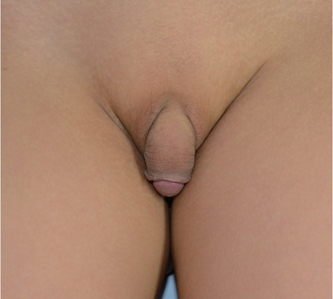
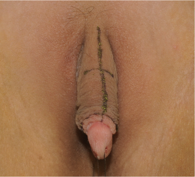
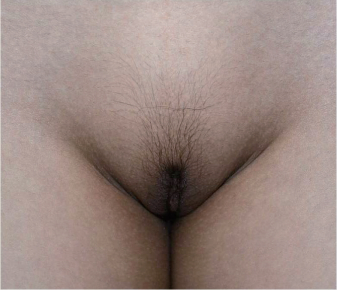
A 6-year-old female presented to our hospital with clitoromegaly. The pediatric department diagnosed the patient with 21-OH deficiency, and she was treated with oral hydrocortisone. There were no complications. On physical examination, she had ambiguous genitalia, precocious pubic hair, and clitoromegaly (Fig. 1). The relaxed clitoris measured 3.7 cm in length. She had grown 16 cm in height over the previous year. Her diagnostic transvaginal ultrasound and abdominal and pelvic computed tomography (CT) showed an invisible uterus with otherwise normal-appearing internal female genitalia. The patient’s karyotype was 46XX. Serum 17-OHP and testosterone levels were increased. We planned reduction clitoroplasty with preservation of the dorsal neurovascular pedicle to maintain the sensitivity of the glans clitoris. First, a 360-degree circumferential incision line 1 cm proximal to the glans sulcus was designed to cover the glans clitoris with the inner mucosa of the prepuce, and a ventral midline incision line was designed (Fig. 2). Next, a stay suture was fixed on the glans. After making an incision along the lines, the corpora cavernosa were dissected from the glans sulcus to the symphysis pubis. Two longitudinal incisions having a wide interval were designed on the Buck’s fascia to include the dorsal neurovascular bundle. The dorsal neurovascular bundle with Buck’s fascia and a strip of tunica albuginea was separated from the corpora cavernosa (Fig. 3A). After that, the corpora cavernosa were completely removed, from the symphysis pubis to the glans sulcus. To reduce the glans size, an incision line was designed on the ventral side of the glans, and reduction of the glans was performed (Fig. 3B). Next, after folding down the dorsal neurovascular bundle, the base of the glans was sutured to the remnant corpora on the symphysis pubis with 4-0 Vicryl. The prepuce of the clitoris was created by trimming the skin, and the rest of the skin was used to reconstruct labia minora. The wound healed well postoperatively. There were no complications over 13 months of follow-up (Fig. 4).
DISCUSSION
Causes of clitoromegaly are divided into hormonal factors such as congenital adrenal hyperplasia and non-hormonal factors such as tumor. Other etiologies include idiopathic causes and pseudoclitoromegaly [3]. Clitoromegaly causes serious psychological stress, and treatment should be performed before 6 to 12 months of age in order to minimize any psychological sequelae [4]. A careful history, physical examination, and hormonal analysis are needed to reach an underlying cause, because clitoromegaly may result from a variety of factors [5]. Techniques for clitoral resection have changed a great deal over the past thirty years as our knowledge of the anatomy has improved. In the past, removal of the whole clitoris (clitorectomy) was used, but is not now an acceptable option because the clitoris is recognized as an important sensory organ involved in sexual response [6]. Later, surgeons tried to preserve as much of the enlarged clitoris as possible and buried the corpora under the skin. However, this meant that the erectile tissue was concertinaed under the skin and, when erect, was very painful [7]. Further modifications of the technique involved excision of corporal tissue and preservation of the glans and the neurovascular bundle to reduce any loss of sensation [8]. On the basis of an extensive study of these techniques, we individualized and combined the most important points of other techniques with our refinements to obtain a better cosmetic result and to preserve clitoral sensitivity. For example, in Papageorgiou et al. [4], both the dorsal and ventral neurovascular bundles were preserved and the glans was untouched. In Kogan et al. [9], the dorsal neurovascular bundle was preserved and the dorsal central wedge glans reduction was performed. However, the surgeon did not reduce the size of the glans clitoris sufficiently, which resulted in an unsightly longitudinal scar on the dorsum of the glans clitoris. Selvaggi et al. [8] removed the glans clitoris bilaterally, but we reduced the glans on the ventral side where the density of nerves is lowest [10]. Ventral glans reduction also has the advantage of hiding the surgical scar and easing the removal of a sufficient amount of tissue. Additionally, we left a wide width of the dorsal neurovascular bundle. This is due to the dorsal nerve, which is widely distributed on the dorsum of the phallus. Finally, in order to cover the glans in a resting state, we constructed a prepuce by trimming the skin. By modifying the various procedures used previously, this operation satisfied both the functional and aesthetic desires of the patient and patient’s parents.