Arch Aesthetic Plast Surg Search

CLOSE


Arch Aesthetic Plast Surg > Volume 20(3); 2014 > Article
Chang, Chen, Cho, and Ahn: Comprehensive Review of Asian Cosmetic Upper Eyelid Oculoplastic Surgery: Asian Blepharoplasty and the Like

Abstract

Background

Asian Aesthetic Oculoplastic Surgery is a fast-growing field, both within the United States and abroad. With growing interest, there have also been multiple terminologies used for the same concepts. This has created redundant and confusing language - prone to errors in patient-physician communication. In addition, there has been an upsurge of various techniques or variations to existing techniques that has created unnecessary confusion among plastic surgeons. The objective of this article is to provide organization and simplification to the terminology and to the techniques used in what some broadly refer to as "Asian Blepharoplasty" or perhaps more correctly termed Asian Aesthetic Oculoplastic Surgery.

Methods

Unified terminology, aesthetic goal and detailed operative technique of commonly conducted Asian blepharoplasty were suggested by experienced oculoplastic surgeons.

Results

The main procedures of Asian Aesthetic Oculoplastic Surgery including supratarsal crease surgery and medial epicanthoplasty were presented with figure and video in this paper. We also have provided author's preferred selection of the major techniques with evaluation of its advantages and disadvantages.

Conclusions

The most important element in patient satisfaction is clear communication of surgical expectations. Then, proper selection of the most suitable pre-operative design, type of surgery performed, and specific crease configuration based on the individual's anatomic and physiological characteristics can be achieved.

INTRODUCTION

The field of Asian Aesthetic Oculoplastic Surgery is a rapidly growing area – with upper eyelid blepharoplasty ranking as the most commonly performed procedure in Asian aesthetic surgery.
The main objective of this article is to provide simplification and reorganization to the surgical procedures of this topic, namely supratarsal crease surgery and epicanthoplasty techniques. In addition, a recommendation for unified terminology and an enhanced explanation of the aesthetic goals of the procedures will be offered. This paper will also evaluate some of the advantages and disadvantages of various surgical approaches in an objective fashion, based on anatomic and physiological criteria of the individual patient at hand.
The imprecise use of terminologies in Asian Aesthetic Oculoplastic Surgery has always been a problem with doctor/patient interaction. This leads to miscommunication, unrealized expectations, and suboptimal results. Because the Asian languages inherently have the capacity to hold multiple meanings even within the same word, there is an even greater importance in using unified language to optimize doctor/patient communication (Table 1).
Too often, complications occur due to faulty primary surgery based on incorrect concepts and choices. Although the choice in terminology may seem to be a minor detail, these are challenging issues when handling patient complaints and suboptimal results in revisional cases. Therefore, it is critical to surgical success to clearly communicate expectations and desires of the patient at the first opportunity.

ANATOMICAL BASIS OF ASIAN AESTHETIC OCULOPLASTIC SURGERY

The anatomical basis of Asian Aesthetic Oculoplastic Surgery revolves around two unique entities, the supratarsal crease and the epicanthal fold. The absence and presence of these two entities distinguishes the aesthetics of the Asian eye from other ethnicities.

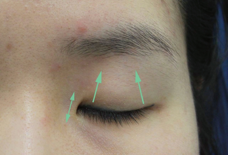
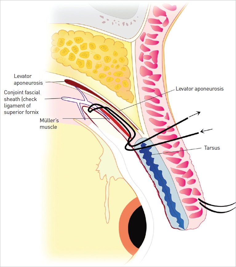
Anatomy and etiology of supratarsal crease

Historically, the most commonly used explanation on the etiology of the supratarsal crease is commonly explained by the Levator Expansion Theory - where the levator aponeurosis sends distal insertions into the overlying dermis of the upper eyelid skin approximately at the level of the superior border of the tarsal plate [1].
However, there have been more recent microanatomic studies that demonstrate how the levator sends distal insertions to overlying orbicularis oculi muscle and subcutaneous tissues, not into the dermis itself [2]. In addition, the general consensus of updated thought is that the contributing factors to the upper lid without a supratarsal crease are the increased thickness of skin, the increase in suborbicularis adipose tissue, the variance in fusional height of the orbital septum and levator aponeurosis, the existence of a pretarsal fat pad, and the increase in preaponeurotic adipose tissues [3-6].

Anatomy and etiology of epicanthal fold

Most Asian epicanthal folds can be categorized as the epicanthus tarsalis variant [7,8]. It is a normal anatomic entity unique to the Asian patient population and is not considered a deformity. The epicanthal fold is comprised of redundant skin, subcutaneous tissue, and a portion of the preseptal orbicularis oculi muscle. When the epicanthal fold is eliminated in the case of epicanthoplasty procedures, there have been no functional deficits found [9].
There have been several anatomical studies published showing the composition of the epicanthal fold [5]. It is located nasally between the upper and lower eyelid, at an epicenter of opposing skin tension vectors. When the upper eyelid opens through the action of the levator muscle and aponeurosis, it is noted that the medial skin of the upper eyelid moves in a superolateral direction and the lateral upper eyelid skin moves in a superomedial direction [10]. (Fig. 1). These vectors of upper eyelid skin tension are more pronounced when the eyelid is open rather than when closed. Additionally, it is prudent to perform an epicanthoplasty procedure concomitantly with a supratarsal crease surgery in order to eliminate and disperse these deterring forces from blunting the supratarsal crease formation.

SUPRATRASAL CREASE SURGERY

The first known published account of the Asian supratarsal crease procedure is in the Japanese literature. In 1896, Mikamo published a suture technique to creating the supratarsal crease [11]. In 1929, Maruo reported the first incisional method [12]. Since then, there have been numerous publications on variations and combinations of these two techniques [13-18], including laser application [19] and the use of microsurgical mini-flaps [20].
There are distinct advantages and disadvantages to each tech nique. Accordingly, one should select the appropriate technique for the individualized patient based on his/her unique anatomic and physiological qualities (eg thin vs thick upper eyelid, age, ocular history, etc.) (Table 2). This is critical to successful surgical outcome and patient satisfaction.
In addition to selecting the appropriate supratarsal crease surgical technique, it is equally important to select the appropriate crease configuration. Clearly establishing the desire crease configuration of the patient is critical to surgical success.

CREASE CONFIGURATION

Supratarsal crease placement - Height

In general, the supratarsal crease should not be placed higher than the superior border of the tarsal plate. This is due to the fact that placement of a crease higher than the superior tarsal border tends to decrease the contractility of the levator muscle and that it is more difficult to lower the crease than to raise it. In addition, a high crease can look unnatural. Unless the patient specifically desires a high placement of the supratarsal crease it is advised to err on the side of conservative, more inferior, placement.

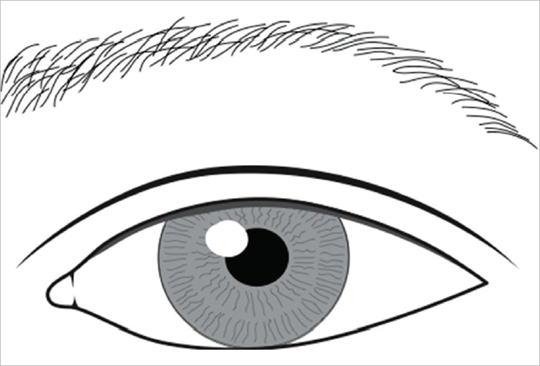
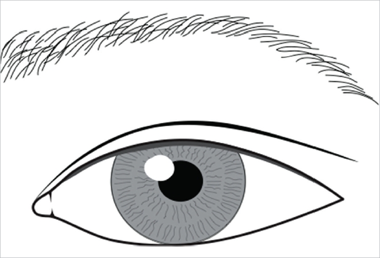
Supratarsal crease configuration - Shape

There are two common crease configurations of the supratarsal crease among Asians: parallel configuration and nasally-tapering configuration (Fig. 2-4). In order to achieve patient satisfaction, there must be a preoperative consensus between patient and surgeon on the selection of the specified crease configuration.

Dynamic crease

The most important aesthetic ideal in the supratarsal crease is the dynamic nature of the crease. The dynamic crease is defined as one that is present when the upper eyelid is open and one that is imperceptible when the eyelid is closed or looking down. To create a supratarsal crease, a component of the anterior lamella (eg dermis) must be fixated to an element of the posterior lamella (eg levator aponeurosis), which is responsible for lid elevation. Whether the incisional technique is utilized or a minimal incision suture technique is utilized, all supratarsal crease surgeries fixate the anterior lamella to the posterior lamella. In order to create a dynamic crease and avoid an uncomely static crease deformity, it is important not to directly anchor the skin to the tarsal plate. The skin flap should instead be fixated to the septoaponeurotic tissues [18,21,22]. This is described in detail in later sections.

Static crease deformity

There are generally two types of static crease deformity: a depressed scar or an overly deep fold. The depressed scar occurs when excessive orbicularis oculi muscle excision was performed – thereby creating a significant muscle gap. The overly-deep fold variant occurs when the lower skin incision flap is anchored too far superiorly onto the tarsus or levator apoenurosis [22].

Aesthetic goals

The aesthetic goals of Asian Oculoplastic surgery are to provide the appearance of a naturally appearing, well-defined, and wideopen palpebral fissure within the normal variations of Asian anatomy. It is important to note that the aesthetic goal of these procedures is not “Westernization” but to create the appearance of a naturalappearing, relatively larger, and open palpebral fissure [23].
The perception of size and shape of the eye is actually based on the perception of the size or shape of the white scleral triangles on either side of the cornea: the nasal scleral triangle and the lateral scleral triangle [26]. The reduction or elimination of the epicanthal fold uncovers, increases, and clearly delineates the nasal scleral triangle - thereby creating the perception of a larger eye. In the case of the supratarsal crease, the presence of a supratarsal crease creates the illusion of a vertically larger palpebral fissure (Fig. 5, 6).
In simple terms, the procedures discussed today – incisional supratarsal crease surgery, minimal-incision supratarsal crease surgery, and epicanthoplasty – all strive to create a larger and naturally appearing palpebral fissure.
In the following section we will discuss the authors’ preferred techniques for Incisional Supratarsal Crease Surgery, Minimal Incision Supratarsal Crease Surgery, and Epicanthoplasty Surgery.

INCISIONAL SUPRATARSAL CREASE SURGICAL TECHNIQUE

Author Chen’s preferred method

The author Chen prefers this incisional method as it provides superior control of crease height and shape to create a permanent, natural crease with lessened incidence of complications. He reports an overall consensus of ~5-10% need for revision among all practitioners of this method. In addition, by fixating the skin to the aponeurotic tissues, this procedure recreates the anatomy thought to be found in naturally-occurring supratarsal creases. This technique also utilizes the same incision for the supratarsal crease creation to reduce the medial canthal upper lid fold, thereby reducing the need for a separate medial epicanthoplasty incision (See below).

Operative technique – Full incision technique [18,27-31]

(Video 1. Incisional Technique)
The appropriate oral premedications (one tablet of Vicodin/hydrocodone acetaminophen as analgesic and 10 milligrams of Valium/diazepam as sedative) are given one hour prior to surgery. Intravenous line is started. The upper lid skin and sub-orbicularis layers are infiltrated with 2% xylocaine with 1:100,000 dilution of epinephrine, along the incision line (A #30 gauge needle is used and the volume injected is seldom over 0.5-0.75 mL for each eyelid). After a 5 minutes period for the anesthetic to disperse, the operative field is prepped and vital signs monitors are applied.
Step 1: Marking of crease incision
Attention is turned to the right eye. A black protective corneal eye shield is applied. The upper lid’s tarsal plate is everted (Fig. 7) and a caliper used to measure the vertical height of the central portion of the tarsal plate. The lid is returned to its normal position and methylene blue ink is used to transpose the measured central tarsal height to the lid skin as the central point of the crease incision(lower line), usually at about 7 mm from the lashes.
Step 2: Skin incision
Depending on clinical finding, typically a segment of skin measuring about 2-3 mm centrally and laterally, and 1 mm medially is included in the upper and lower lines of incision (Fig. 8). Skin incision is made using a #15 blade.
Step 3: Beveled transection through orbicularis oculi layer[27]
When the orbicularis muscle is seen, control of capillary oozing is carried out using bipolar cautery. This layer is carefully traversed using surgical blade or monopolar cautery with cutting mode on a low energy setting. The cautery tip is intentionally beveled superiorly along the orbicularis such that the orbital septum is reached at a slightly higher level from the superior tarsal border. The beveled approach allows a safer route to the preaponeurotic space while avoiding inadvertent touch of the levator muscle.
Step 4: Opening of orbital septum
When the preaponeurotic fat is seen through a small opening of the septum, the septum is opened horizontally with a blunt scissors (Westcott’s), avoiding blood vessels in the fat pads or the levator aponeurosis underneath.
Step 5: Treatment of fat pads
The strip of skin-muscle bounded by the two skin incisions, is retracted inferiorly using a Blair retractor. If preaponeurotic fat is prolapsing significantly down and overlaps the superior tarsal border where the crease construction is to occur, it can be partially excised.
Step 6: Trapezoidal debulking and trimming of the skin-muscle strip along the lower skin incision at level of superior tarsal border (Fig. 9)
The preaponeurotic platform has been cleared for optimal interplay between the posterior lamella of levator and Mueller’s muscle with its attached tarsus, and the relaxed anterior skin and orbicularis oculi; there is now less hindrance (impedance) to dynamic crease construction. The crease forms well even without wound closure at this point.
Step 7: Resetting of tissue plane
At this point, it is essential to release the forehead drapes and properly reposition the brow and upper lid tissues in relation to the underlying levator aponeurosis and lower pretarsal tissues. It avoids setting an exaggerated crease height, induction of secondary ptosis, lagopthalmos on downgaze, and a secondary higharched brow as a compensatory reaction. This is a most under-appreciated step in upper blepharoplasty. In revisional cases, this resetting allows some skin recruitment and brings in additional soft tissues to partially fill in any deep sulcus from fat excision associated with previous blepharoplasty.
At this point is where management of medial upper lid fold comes in: Often among patients who present a clinically evident medial upper lid fold, there may be a need to reduce the prominence of this fold. The author uses the following technique as an added solution set:
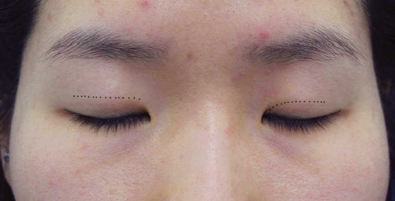
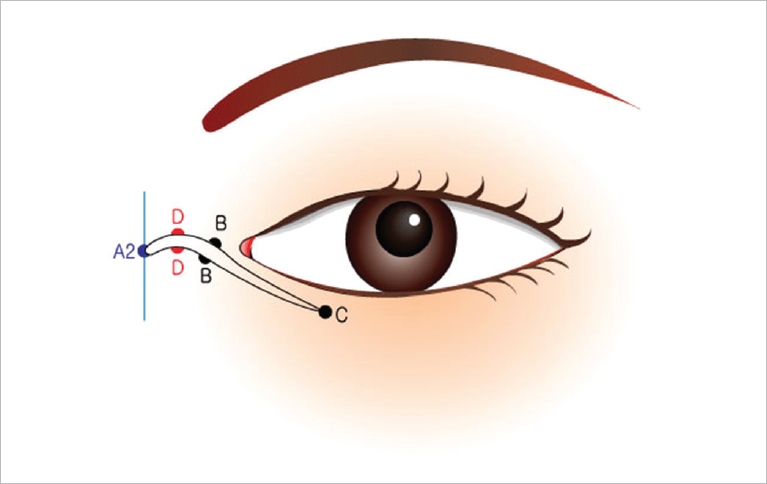
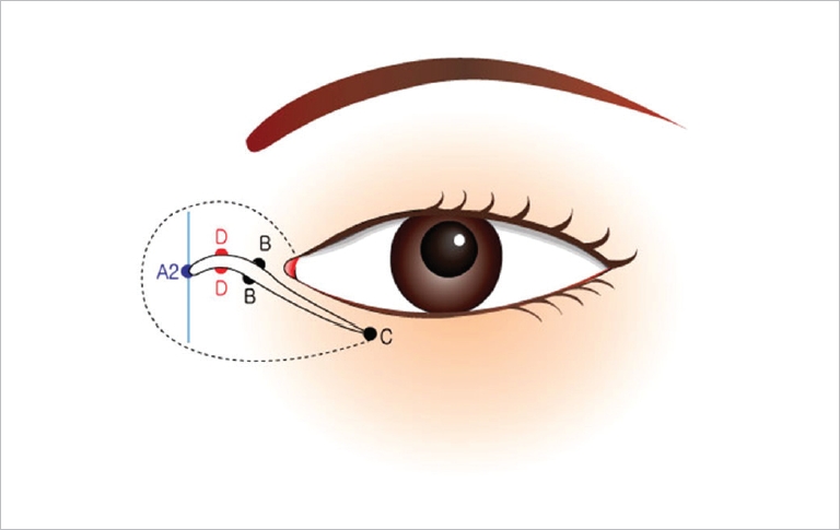
  • (i) In the design of the nasally-tapered crease, the incision lines are drawn to converge towards the medial canthus as well as the medial fold. To enhance the crease invagination to blend in under the fold, the area is slightly undermined, and excess skin tissue is cut just below the converging crease line (infracrease cut, first snip); the lower edge’s skin is migrated up and a second cut takes care of the small dog-ear. The crease is closed with an interrupted suture placed with the knot on the lower side of the wound edge (infraanchoring of knot,“down-knot” the crease). The goal is to have the nasal-tapering crease indent to beneath (inferior to) the natural residual fold. (Alternatively one may also make the first snip along the crest of the medial canthal fold, and then migrate the lower edge’s excess skin upward to trim it; then infra-anchor its closing knot. It has a slightly higher chance of postoperative induration since the skin along the crest of the medial fold is slightly thicker.)

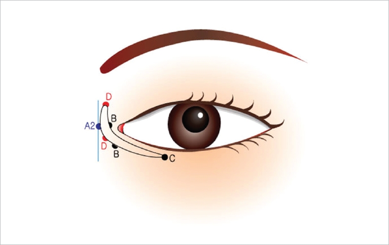
  • (ii) With a parallel shape design, after the main steps are carried out and the tissue planes have been reset, the area under the medial fold is slightly undermined. The first relaxing snip near the medial end of the undermined redundant skin is biased towards the lower incision in a slightly oblique fashion (infra-crease cut); the lower edge’s excess skin is migrated upward such that the medial fold is reduced with the second cut removing the dog ear and based along the medial direction of the parallel crease shape. The closure at this end is with a fine interrupted suture tied very lightly and laid on the superior side of the incision (supra-placement of knot without anchoring; “up-knot” the tie loosely, the rationale being that the medial end of a natural parallel crease tends to be shallow). These delicate techniques I used have handled the ethnic non-pathologic medial upper lid fold quite well without the complicated steps usually associated with treatment of epicanthal fold (Epicanthus), which is usually reserved for findings in Down’s syndrome, or patients born with blepharophimosis [32].

Step 8: Crease construction and wound closure
#6-0 sutures are applied from the lower skin edge, picking up small strands of the levator aponeurosis just above the superior tarsal border, then through the upper skin edge and then tied down. Six to nine interrupted sutures are typically used. The rest of the skin gap is closed using 7-0 prolene, nylon or silk (Fig. 10C). The eye shield is removed from the right eye and applied to the left eye where the procedure is repeated. When completed, each eye is flushed with normal saline. The surgeon checks for symmetry in the crease and makes necessary adjustment.

Post-operative management

The patient receives ice compresses to the upper eyelids and is instructed to continue icing and bed rest for one day. The patient is told to have restricted physical exertion the first week. Topical tobramycin or gentamicin-prednisolone ophthalmic ointment (PredG, by Allergan) is applied four times daily for one week. Suture removal is usually in one week. Fig. 11 shows three female patients before and at two months postoperatively; with the first patient’s crease height set at a slightly below-average crease height of 6.5 mm, the middle set is at medium-average crease height of 7.0 mm, while the right set is at higher than average crease height of 7.5 mm.

MINIMAL INCISION SUPRATARSAL CREASE SURGERY

Author Ahn’s preferred minimal incision method - triangular 5-microincision suture method

The author Ahn prefers this method over the classic rectangular 3-microincisional suture technique as the additional micro-incisions and triangular configuration creates a more even crease, increases the fixation of anterior to posterior lamella, and decreases the overall area of potential tissue strangulation with triangles over rectangles (Fig. 12). Of note, addition of a Muller-muscle loop plication to this technique can create a simultaneous mild ptosis correction [17] (Fig. 13).

Operative technique – 5 point triangular continuous suture technique [17]

(Video 2. Minimal Incision – 5 Point Triangular Continuous Suture Technique)
Step 1: Preoperative markings are made with the patient in sitting upright position
It is critical to determine whether the patient desires a parallel or a nasally-tapering crease configuration.
Step 2: The patient is brought to the operating room and placed in supine position
The procedure is performed under conscious sedation and local anesthetics. After reconfirmation of all markings, the anterior lamella and posterior lamella is injected with a half-inch #26 gauge needle containing 2% lidocaine and 1:50,000 epinephrine. Anesthetic eye-drops are placed as well.
Step 3: Using a #11 scalpel tip, five micro-incisions are made at equal intervals just above the level of the superior border of the tarsal plate. Number the slits from 1 through 5, starting medially and ending laterally. Use iris scissors to dissect through the micro-incisions for a larger opening subdermally. By utilizing a lateral microincision, the risk of inadvertent levator injury is decreased. Through the 4th microincision, grab the orbicularis oculi muscle with 0.5 forceps and cauterize the muscle carefully through the forceps. Do not allow the forceps to come in contact with the skin. (This allows access to the post-septal region for debulking the thicker-lidded patient.)
Step 4: In those patients with thicker lids, carefully grab the preaponeurotic fat through the 4th micro-incision. Through gentle pulling and teasing motions, carefully deliver a conservative amount of adipose tissue to be removed. Using a mosquito clamp, carefully clamp the adipose tissue, cut the adipose tissue, and cauterize the stump before removal of the clamp. Allow the cauterized adipose stump to retract back into the lid.
Step 5: Utilize corneal protection with either a corneal shield or use of a Jaeger lid plate. The suture used is a #7-0 blue nylon on a long tapered needle. The suture is introduced through the 4th slit subcutaneously and passed out through the 3rd slit. From the 3rd slit, the suture is passed posteriorly through the conjunctiva from the everted lid right above the superior edge of the tarsal plate (~1-2 mm). Then, the suture is passed back out from the same point in the conjunctiva out of the 2nd skin micro-incision. Next, pass the suture subcutaneously from the 2nd to the 1st skin micro-incision. The suture is then passed again back through the conjunctiva of the everted lid right above the superior edge of the tarsal plate. Then, the suture is passed back out from this point to the 2nd skin microincision. Repeat this pattern back to the 4th skin micro-incision, utilizing all 5 micro-incisions. This completes triangular-shaped continuous suture path (Fig. 12).
Step 6: When the triangular-shaped continuous suture run is complete, the suture should be exiting the same hole that the suture was first introduced, the 4th microincision. At this point, tie the knot over a blunt needle or a cotton-tip at the appropriate tension to avoid purse-stringing and strangulation of the lid tissues. Cut the knot and allow knot to retract into the lid. Note, over-tightening the knot can cause increased tissue strangulation when post-operative edema sets in.
Step 7: Repeat on the contralateral lid.
Step 8: If the patient has normal levator function and mild ptosis (1-2 mm), a Müller muscle plication loop can be added during the triangular-shaped continuous suture path (between the 3rd and 4th micro-incision in the conjunctiva side) by plicating the Müller’s muscle subconjunctivally from the level at the superior border of the tarsal plate to approximately 10-15 mm cephalad (average is between 12-13 mm) towards the superior sulcus and back to the superior border of the tarsal plate before passing the suture back out of the 4th skin micro-incision (Fig. 13).

Postoperative management

Instruct patient to maintain head elevation when possible and to use ice for comfort as needed.

EPICANTHOPLASTY

General overview/patient selection

In many publications, measurements of various periorbital values are used to determine whether the epicanthoplasty procedure should be performed. These include values such as interepicanthal distance, intercanthal distance, and horizontal palpebral fissure dimensions [33,34]. At times, these calculations can be complicated and confusing. It is simple to note that the main aesthetic effect of the epicanthoplasty procedure is that of enlarging the nasal scleral triangle and creating the perception of a larger palpebral fissure. Also, in the patient with an epicanthal fold, performing the supratarsal crease procedure alone diminishes its effect by leaving the blunted nasal scleral triangle made by the persistent epicanthal fold.
Generally speaking, in the Asian patient who desires a paralleloriented crease, it is aesthetically pleasing to have a small or nonexistent epicanthal fold. Also, in those patients with very large caruncles, it might be advised not to perform epicanthoplasty to keep the caruncle from over-exposure.

Skin-Redraping Epicanthoplasty [33]

There are multitude of published epicanthoplasty techniques ranging from simple direct excision, Z-plasty, Y-V advancement method, W-plasty, etc. Among those, the more popular techniques are the Uchida split V-W technique [7,35], modified Mustarde technique [36], and the half Z-plasty technique. Recently, the skinredraping technique has become one of the more popular techniques. [33]

Operative technique – Skin redraping technique

(Video 3. Epicanthoplasty – Skin-Redraping Technique)
Step 1: With the patient sitting upright and in primary gaze, use a finger to retract the epicanthal fold in a horizontal vector towards the nasal direction. With the medial canthus/caruncle uncovered by the retracted epicanthal fold, mark a dot (B) 2 mm nasal to the lacrimal lake of the medial canthal region (Fig. 14).
Step 2: Then lift the marker off of point B and while holding the marker in place in the air, release the digitally retracted epicanthal fold and place a mark (A1) where the marker lands (superimposed point). (Of note, point B and A1 should lie along the same horizontal axis parallel to the floor) (Fig. 15).
Step 3: Next, draw a dotted line that is perpendicular to this horizontal axis at point A1. At a point that is approximately 70-80% of the distance between A1 and B, mark point A2. (Of note, point A2 should be along the same horizontal axis as points A1 and B). The distance from A1 to A2 in relationship to the distance from A2 to B should be a 2:8 ratio (Fig. 16).
Step 4: Next, draw a solid line that is perpendicular to the horizontal axis through point A2. At a point that is nasal to the medial limbus of the iris, mark point C along the subciliary line. Now, mark point D between A2 and B but at a location slightly higher/cephalad than the horizontal axis where point A1, A2 and B lay. Note, the incision line will be from point A2, through point D, point B, and ends at point C (Fig. 16).
Step 5: After these preoperative markings are made, the area is infiltrated with local anesthesia.
Step 6: Incision is made with a #15 scalpel through skin and subcutaneous tissues along the marked incision line (Fig. 17).
Step 7: Undermining is done superior (cephalad) and inferiorly (caudad) to the incision line (Fig. 18).
Step 8: The orbicularis muscle of the epicanthal fold is transected sharply and trimmed. Once this is done, meticulous hemostasis is achieved. The skin will now redrape into the configuration shown in Fig. 19. The inferior skin flap will have a tendency to move more laterally and the superior skin flap will have a tendency to move in a more cephalad direction. As you start closure of the skin incision by closing point A2 to the upper skin flap’s B point, a dog-ear will form around the upper skin flap’s point D. This dog-ear can be directly excised or can be treated with laser resurfacing. In addition, during closure, a length discrepancy will be noted between the two skin edges. It is important to redistribute the skin incision flaps evenly to evenly disperse and eliminate this length discrepancy during meticulous closure.
If an epicanthoplasty is performed with the primary intent to relieve the tension created by the fold, use a much shorter incision where point A2 is halfway or less between point A1 and point B (60:40 ratio). On the reverse side, if point A2 is made at a point larger than 70-80% of the distance between A1 and B (e.g. 1:9), there will be a great overcorrection and the patient will have a stretched and pronounced caruncular show.

Postoperative management

In normal postoperative course, most epicanthoplasty scars will mature to imperceptible state with at least 3 months time. In the case where there appears to be hypertrophic scarring after this time, adjuvant CO2 laser resurfacing at the site can improve the scarring. Some surgeons advocate steroid micro-injection at the time of surgery and postoperatively as well. With proper preoperative counseling of expected 3 month course of healing, most patient expectations can be managed.

CONCLUSIONS

This paper has highlighted the need for unified terminology in the field of Asian Aesthetic Oculoplastic Surgery. In addition, we have re-examined and simplified the aesthetic goals of the main procedures. We have provided author’s preferred selection of the major techniques with evaluation of its advantages and disadvantages.
In conclusion, the most important element in patient satisfaction is clear communication of surgical expectations. Then, proper selection of the most suitable pre-operative design, type of surgery performed, and specific crease configuration based on the individual’s anatomic and physiological characteristics can be achieved.

CONFLICTS OF INTEREST

None of the authors has a financial interest in any of the products, devices, or drugs mentioned in this manuscript.

Fig. 1.
Skin tension vectors.
aaps-2014-20-3-129f1.jpg
Fig. 2.
Parallel configuration.
aaps-2014-20-3-129f2.jpg
Fig. 3.
Nasally-tapered configuration.
aaps-2014-20-3-129f3.jpg
Fig. 4.
Parallel vs nasally-tapering supratarsal crease design.
aaps-2014-20-3-129f4.jpg
Fig. 5.
Absence of supratarsal crease.
aaps-2014-20-3-129f5.jpg
Fig. 6.
Presence of supratarsal crease – after supratarsal crease surgery.
aaps-2014-20-3-129f6.jpg
Fig. 7.
Everted Lid. Right upper lid is everted and the height of the tarsus is measured centrally with a caliper from the posterior lid margin to the superior tarsal border.
aaps-2014-20-3-129f7.jpg
Fig. 8.
Incision. Skin incision has been made, first along the lower line of incision - which will become the new crease line, and then along the upper line of incision.
aaps-2014-20-3-129f8.jpg
Fig. 9.
Preaponeurotic space. Preaponeurotic space and Trapezoidal debulking and trimming of the skin-muscle strip – This maneuver often facilitates the glide mechanism [28] (glide plane) of the levator relative to the anterior skin/orbicularis oculi layer, allowing an eyelid crease pulled by the levator/Muller’s to indent dynamically against a relaxed preseptal skin-orbicularis layer which forms the eyelid fold.
aaps-2014-20-3-129f9.jpg
Fig. 10.
Crease construction and wound closure. (A) procedure completed – eyelid in passive repose. (B) Upper lid opened, crease indents naturally prior to closure without any skin stitched. (C) Completion of wound closure, the lid opens freely with good crease indentation.
aaps-2014-20-3-129f10.jpg
Fig. 11.
Pre-operative and post-operative photos.
aaps-2014-20-3-129f11.jpg
Fig. 12.
The 5-point triangular continuous suture technique.
aaps-2014-20-3-129f12.jpg
Fig. 13.
Coronal section of the 5-Point Triangular Continuous Suture Technique with Muller-Muscle Loop Plication.
aaps-2014-20-3-129f13.jpg
Fig. 14.
With the patient sitting upright and in primary gaze, use a finger to retract the epicanthal fold in a horizontal vector towards the nasal direction. With the medial canthus/caruncle uncovered by the retracted epicanthal fold, mark a dot (B) 2 mm nasal to the lacrimal lake of the medial canthal region.
aaps-2014-20-3-129f14.jpg
Fig. 15.
Then lift the marker off of point B and while holding the marker in place in the air, release the digitally retracted epicanthal fold and place a mark (A1) where the marker lands (superimposed point).
aaps-2014-20-3-129f15.jpg
Fig. 16.
At a point that is approximately 70-80% of the distance between A1 and B, mark point A2. (Of note, point A2 should be along the same horizontal axis as points A1 and B).
aaps-2014-20-3-129f16.jpg
Fig. 17.
Incision is made with a #15 scalpel through skin and subcutaneous tissues along the marked incision line.
aaps-2014-20-3-129f17.jpg
Fig. 18.
Undermining is done superior (cephalad) and inferiorly (caudad) to the incision line.
aaps-2014-20-3-129f18.jpg
Fig. 19.
The skin will now redrape into the configuration shown in this Figure.
aaps-2014-20-3-129f19.jpg
Table 1.
Terminology in Asian aesthetic oculoplastic surgery
Identity Current common terms Proposed term – Term used in this manuscript
Supratarsal crease creation surgery Asian blepharoplasty, double eyelid surgery, lid crease surgery, Asian upper lid procedure Supratarsal crease surgery
Parallel oriented supratarsal crease “Outfold”, “outer-oriented” crease, parallel crease Parallel crease
Nasally tapered supratarsal crease “Infold”, “inner-oriented” crease, “unfolded fan” fold Nasally tapered crease
Epicanthal fold “Mongolian fold” Epicanthal fold
Hypertrophic orbicularis roll along lower lash line “Love-roll”
Microincisional buried suture technique Non-incisional, suture technique Minimal incision supratarsal crease surgery
Table 2.
Incisional vs minimal incision techniques in supratarsal crease surgery
Incisional Minimal incision
Ideal patient All patients, especially useful in revision cases, thick-lid cases, or patients with dermatochalasis Thin-lid patient
Advantages Direct visualization, can perform ptosis correction concomitantly, ability to perform selective tissue debulking, Better ability to control for hemostasis, Previse preservation/repositioning of preapoenurotic fat Minimal external scarring, shorter recovery time
Disadvantages Longer recovery time, possibly greater learning curve Decreased visualization of tissues, more difficulty in creating a parallel-oriented crease (with disappearance of medial crease), permanent buried sutures placed, potential Faden Effect [30]
Complications Possibility of static crease deformity, asymmetry Suture failure (recurrence), suture erosion, suture palpability, asymmetry, partial crease formation, feeling of “strain” in eyelids

REFERENCES

1. Sayoc BT. Absence of superior palpebral fold in slit eyes; an anatomic and physiologic explanation. Am J Ophthalmol 1956;42:298-300.
crossref pmid
2. Morikawa K, Yamamoto H, Uchinuma E, et al. Scanning electron microscopic study on double and single eyelids in Orientals. Aesthetic Plast Surg 2001;25:20-4.
crossref pmid
3. Jeong S, Lemke BN, Dortzbach RK, et al. The Asian upper eyelid: an anatomical study with comparison to the caucasian eyelid. Arch Ophthalmol 1999;117:907-12.
crossref pmid
4. Cheng J, Xu FZ. Anatomic microstructure of the upper eyelid in the Oriental double eyelid. Plast Reconstr Surg 2001;107:1665-8.
crossref pmid
5. Kakizaki H, Takahasi Y, Nakano T, et al. The causative factors or characteristics of the Asian double eyelid: an anatomic study. Ophthal Plast Reconstr Surg 2012;28:376-81.
crossref pmid
6. Bang YH, Chu HH, Park SH, et al. The fallacy of the levator expansion theory. Plast Reconstr Surg 1999;103:1788-91.
crossref pmid
7. Park JI. Z-Epicanthoplasty in Asian Eyelids. Plast Reconstr Surg 1996;98:602-9.
crossref pmid
8. Yoo WM, Park SH, Kwag DR. Root Z-epicanthoplasty in Asian eyelids. Plast Reconstr Surg 2002;109:2067-71.
crossref pmid
9. Lee Y, Lee E, Park WJ. Anchor epicanthoplasty combined with outfold type double eyelidplasty for Asians: do we have to make an additional scar to correct the Asian epicanthal fold? Plast Reconstr Surg 2000;105:1872-80.
crossref pmid
10. Kakizaki H, Zako M, Ide A, et al. Causes of undercorrection of medial palpebral fissures in blepharoptosis surgery. Ophthal Plast Reconstr Surg 2004;20:198-201.
crossref pmid
11. Mikamo M. Mikamo’s double-eyelid operation: the advent of Japanese aesthetic surgery. 1896. Plast Reconstr Surg 1997;99:664.
pmid
12. Shirakabe Y, Kinugasa T, Kawata M, et al. The double eyelid operation in Japan: its evolution as related to cultural changes. Ann Plast Surg 1985;15:224-41.
crossref pmid
13. Cho BC, Byun JS. New technique combined with suture and incision method for creating a more physiologically natural double-eyelid. Plast Reconstr Surg 2010;125:324-31.
crossref pmid
14. Liu X, Fan D, Guo X, et al. A transcutaneous, subcutaneous, and intratarsal suturing procedure in double eyelid surgery. Plast Reconstr Surg 2010;126:2133-9.
crossref pmid
15. Baek SM, Kim SS, Tokunaga S, et al. Oriental blepharoplasty: single stitch, non-incision technique. Plast Reconstr Surg 1989;83:236-42.
crossref pmid
16. Mutou Y, Mutou H. Intradermal double eyelid operation and its follow-up results. Br J Plast Surg 1972;25:285-91.
crossref pmid
17. Ahn TJ, Kim KK. Cases of mild ptosis correction with suture-method. Arch of Aesthetic Plast Surg 2012;18:15-20.

18. Chen WP. Asian blepharoplasty. Update on anatomy and techniques. Ophthal Plas Reconstr Surg 1987;3:135-40.
crossref
19. Kang DH, Koo SH, Choi JH, et al. Laser blepharoplasty for making double eyelids in Asians. Plast Reconstr Surg 2001;107:1884-9.
crossref pmid
20. Choi Y, Eo S. A new crease fixation technique for double eyelidplasty using mini-flaps derived from pretarsal levator tissues. Plast Reconstr Surg 2010;126:1048-57.
crossref pmid
21. Kim HS, Hwang K, Kim CK, et al. Double-eyelid surgery using septoaponeurosisj junctional thickening results in dynamic fold in Asians. Plast Reconstr Surg Glob Open 2013;1:e14.

22. Cho IC. The Art of Blepharoplasty. Seoul: Koonja; 2013.

23. Nguyen HN, Nguyen N. P92:Upper blepharoplasty for Asian eyelid is not a westernizing procedure. Plast Reconstr Surg 2006;118 Supplement 4:172.
crossref
24. Price KM, Gupta PK, Woodward JA, et al. Eyebrow and eyelid dimensions: an anthropometric analysis of African Americans and Caucasians. Plast Reconstr Surg 2009;124:615-23.
crossref pmid
25. Park DH, Choi WS, Yoon SH, et al. Anthropometry of Asian eyelids by age. Plast Reconstr Surg 2008;121:1405-13.
crossref pmid
26. McCord CD, Codner MA. Eyelid and Periorbital Surgery. St. Louis: Quality Medical Publishing; 2008.

27. Chen WP. Concept of triangular, trapezoidal and rectangular debulking of eyelid tissues: application in Asian blepharoplasty. Plast Reconstr Surg 1996;97:212-8.
crossref pmid
28. Chen WP. The concept of a glide zone as it relates to upper lid crease, lid fold, and application in upper blepharoplasty. Plast Reconstr Surg 2007;119:379-86.
crossref pmid
29. Chen WP. Beveled approach for revisional surgery in Asian blepharoplasty. Plast Reconstr Surg 2007;120:545-52.
crossref pmid
30. Chen WP, Chen KL. The Eyelid Crease and Double Eyelid Surgery. Cupertino: iBooks textbook; 2012.

31. Chen WP, Khan J. Color Atlas of Cosmetic Oculofacial Surgery. Philadelphia: Elsevier Saunders; 2010.

32. Johnson CC. Epicanthus. Am J Ophthalmol 1968;66:939-46.
crossref pmid
33. Oh YW, Seul CH, Yoo WM. Medial epicanthoplasty using the skin redraping method. Plast Reconstr Surg 2007;119:703-10.
crossref pmid
34. Fujiwara T, Maeda M, Kuwae K, et al. Modified split V-W Plasty for entropion with an epicanthal fold in Asian eyelids. Plast Reconstr Surg 2006;118:635-42.
crossref pmid
35. Ahn SY. Epicanthoplasty with modified Uchida method. J Korean Soc Aesth Plast Reconstr Surg 2002;8:75-80.

36. Yoon K. Modification of Mustardé technique for correction of epicanthus in Asian patients. Plast Reconstr Surg 1993;92:1182-6.
crossref pmid
TOOLS
Share :
Facebook Twitter Linked In Google+ Line it
METRICS Graph View
  • 12 Crossref
  •    
  • 11,360 View
  • 435 Download
Related articles in AAPS


ABOUT
ARTICLE & TOPICS
Article Category

Browse all articles >

TOPICS

Browse all articles >

BROWSE ARTICLES
AUTHOR INFORMATION
Editorial Office
101-2003, Lotte Castle President 109 Mapo-daero, Mapo-gu, Seoul 04146, Korea
Tel: +82-2-3472-4243    Fax: +82-2-3472-4254    E-mail: ksaps@ksaps.or.kr                

Copyright © 2024 by Korean Society for Aesthetic Plastic Surgery.

Developed in M2PI

Close layer
prev next