INTRODUCTION
Trigeminal trophic syndrome (TTS) is a rare disease that presents with paresthesia, anesthesia, and facial ulceration after trigeminal nerve injury. Its main causes are stroke or trigeminal nerve ablation, although it can rarely be caused by infectious agents such as herpes zoster or mycobacteria [1]. TTS tends not to heal well and recurs frequently. The anatomical location of the ala nasi also further complicates treatment.
There is no established treatment protocol for TTS, and limited treatment modalities have been attempted in the past. Wound dressing and medical treatments for paresthesia have been used as an option, but more elaborate surgical methods such as skin grafting and flap placement may be required in severe cases. In this regard, plastic surgeons play a very important role in the treatment of TTS.
Although attempts have been made to use local flaps in cases of TTS, they were not able to cover large defects. Recurrence was also frequent, as local flaps used the affected skin adjacent to the lesion [2]. Thereafter, regional flaps were attempted, and successful treatment of TTS on the ala nasi using a forehead flap was reported [3]. However, forehead flaps cause cosmetic problems due to donor site scarring and their bulky size. Therefore, another method was needed to address these problems.
Herein, we report, for the first time, a rare case of TTS occurring after herpes zoster infection that was successfully treated with a folded nasolabial flap. Cosmetic results were excellent without nostril collapse, extensive scarring of the donor site, or relapse. This rare case could be considered educational for plastic surgeons.
CASE REPORT
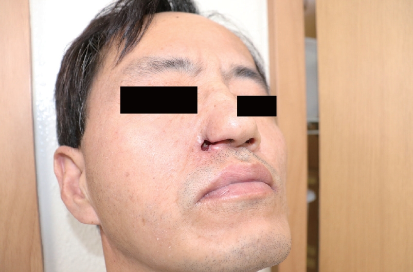
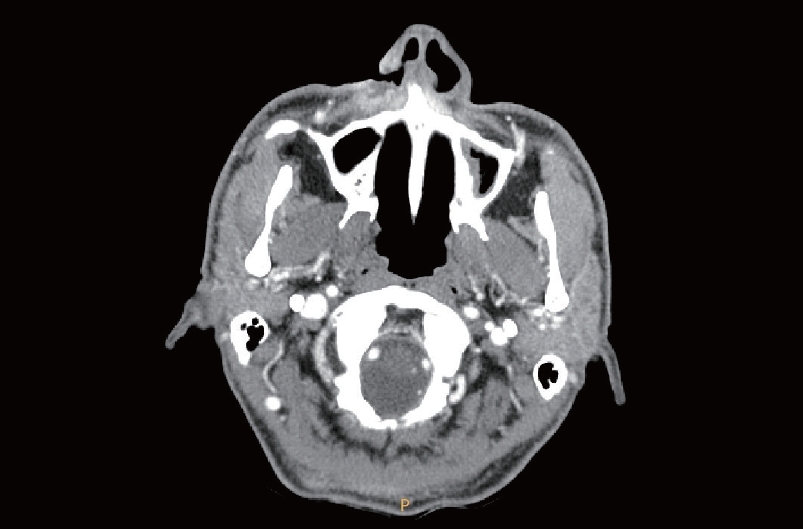
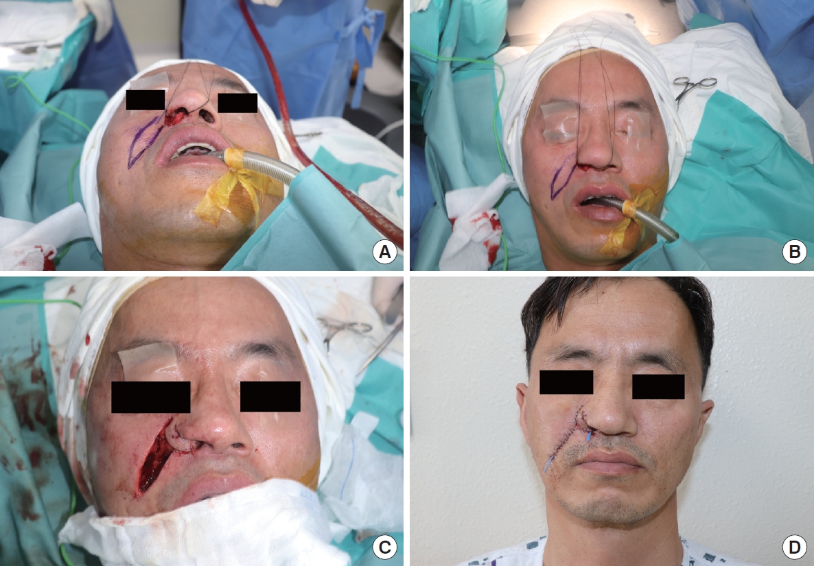
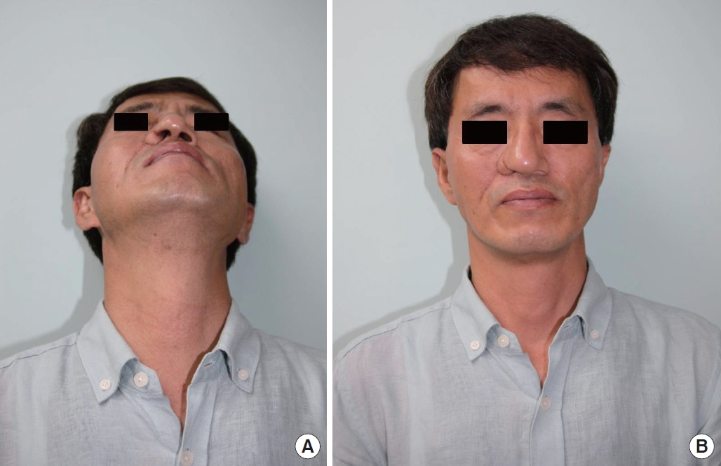
A 51-year-old man was referred to the department of plastic and reconstructive surgery for a skin defect on the right ala nasi. Three weeks prior, he had presented with painful vesicles on the right V2 dermatome with edema. He was diagnosed with herpes zoster and hospitalized for intravenous antiviral therapy. After 1 week of treatment, the vesicular lesions and symptoms were relieved. However, a skin defect developed on the right ala after discharge, and he complained of paresthesia and a tingling sensation at the defect. Clinically, a 1.5├Ś1 cm full-thickness skin defect was observed in the lateral portion of the right ala, including the alar rim. The defect was accompanied by partial loss of the lower lateral cartilage and nostril collapse (Fig. 1). Facial computed tomography revealed a soft tissue defect in the right ala with septal deviation (Fig. 2). The surrounding tissue showed abnormal enhancement and abscess formation. Excisional biopsy was performed to rule out skin defects due to skin malignancy and mycobacterial or fungal infections. Histologically, inflamed granulation tissue was found, and immunohistochemical staining was negative for periodic acid-Schiff and GrocottŌĆÖs methenamine silver stain. Tissue cultures for tuberculosis and fungi showed no growth. As the patientŌĆÖs symptoms occurred after a trigeminal nerve insult, he was diagnosed with TTS induced by herpes zoster infection and hospitalized for reconstructive surgery of the lesion. A two-stage folded nasolabial flap was planned. Margin trimming was performed under general anesthesia, and a folded nasolabial flap was designed to cover both the outer and inner linings of the full-thickness defect (Fig. 3A and B). The angular artery was identified and used as the flap pedicle. The flap was elevated, with the proximal end covering the outer lining of the defect (Fig. 3C). The distal end was folded without distortion to cover the inner lining of the defect. The donor site was closed using a primary suture (Fig. 3D). Although the flap was slightly bulky immediately after the surgery, it was relatively symmetrical and well-supported, despite alar reconstruction having been performed with only a skin flap and no bony framework such as cartilage or implant. The flap was well maintained without nose collapse, and there was no evidence of recurrence at a 1-year follow-up (Fig. 4). The patient was scheduled for secondary surgery for division and contouring in the future.
DISCUSSION
TTS is a rare skin condition first described by Loveman in 1993, characterized by neurotrophic ulceration of the face, especially the ala nasi, occurring after trigeminal nerve injury [4,5]. It is accompanied by paresthesia, such as burning or tingling, and pain perception is reduced or absent. Severe ulceration may cause skin defects, resulting in persistent cosmetic sequelae, such as extensive scars. It has been reported to occur more often in women and mainly affects the right face. Involvement of facial areas other than the nose, such as the cheek, lips, scalp, forehead, or even the bilateral face, has been reported in rare cases [6,7].
TTS needs to be clinically differentiated from other diseases that present with persistent or chronic ulcerations. Although the histological features of TTS are nonspecific, skin biopsy may be required to rule out skin malignancies such as basal cell carcinoma and squamous cell carcinoma. It is also important to differentiate TTS from infections caused by fungi, mycobacteria, syphilis, and parasites. Culture or immunohistochemical staining through skin swabs or tissue biopsy should therefore be performed. In our case, skin biopsy, tissue culture, and staining were performed to rule out the differential diagnoses mentioned above. The patient was finally diagnosed with TTS because no evidence of malignancy or infection was found.
The causes of trigeminal nerve damage that can induce TTS are diverse. Stroke, including WallenbergŌĆÖs syndrome, which causes posterior inferior cerebellar artery occlusion, is one cause. In addition, nerve injury due to trauma or trigeminal nerve ablation such as rhizotomy or nerve block for the treatment of trigeminal nerve neuralgia can also induce TTS. In rare cases, TTS can result from infections such as herpes zoster or leprosy [1]. The period between trigeminal nerve injury and ulcer development can vary from several weeks to years, with an average of 2 years [8]. In our case, ulceration of the ala nasi occurred 3 weeks after herpes zoster infection in the V2 dermatome.
TTS shows a persistent pattern after it occurs, making it difficult to treat. Various methods have been proposed for this purpose. Dressing methods with hydrocolloid, occlusive materials, or topical antibiotics have been attempted. Successful treatment of TTS using negative pressure wound therapy with a vacuum device has also been reported [9]. Transcutaneous electrical nerve stimulation and medication therapy with carbamazepine, amitriptyline, or diazepam have been administered for paresthesia and neuralgia [8]. In case of deep ulcerations or full-thickness skin defects that do not respond to these treatments, surgical treatment such as a skin graft or flap coverage can be attempted [10].
The surgical treatment of TTS is difficult because of subsequent contractions, poor support of the nostrils, and high recurrence rates. Local flaps donated from the affected area of the face can lead to poor results [2]. In addition, local flaps are not useful for large or full-thickness skin defects, so regional flaps have been attempted to treat TTS. The forehead flap and septal mucoperichondrial flap are conventional reconstructive options for full-thickness alar defects [11]. In particular, the forehead flap has been widely used to reconstruct defects of all nose subunits of a wide range of sizes [12]. However, it is technically challenging to create asymmetry and to minimize scarring. A previous case report of TTS treated with a pedicled forehead flap stated that the flap was well-maintained without nostril collapse or recurrence [3]. However, the forehead flap induces cosmetic problems due to its bulky size and severe scarring of the donor site. The septal mucoperichondrial flap is a type of lining flap, and a thinner nostril lining can be expected with this technique than with other local flaps. However, it requires a high level of technical skill, making it difficult to accurately predict surgical outcomes.
Therefore, in our case, a folded nasolabial fold flap was used in an attempt to overcome the disadvantages of the forehead flap and septal mucoperichondrial flap. Since this flap is designed parallel to the nasolabial fold, it has better cosmetic outcomes in terms of reasonable flap size and less donor site scarring. In addition, the procedure is simpler, so it requires a shorter operation time than the other methods.
TTS is a rare and challenging skin condition encountered by plastic surgeons. When encountering a combination of anesthesia, paresthesia, and ulceration, physicians should suspect TTS. It is necessary to take a thorough history to determine the cause of trigeminal nerve injury that induced TTS. Tissue culture and biopsy may be performed to rule out ulcerative skin malignancy or fungal/mycobacterial infection. A dressing can be attempted, but a surgical intervention is recommended for extensive ulceration or full-thickness skin defects. A regional flap is more effective than a local flap because of the possibility of the skin around the ulceration also being affected. To our knowledge, there have been no attempts to use folded nasolabial flaps for the treatment of TTS. Therefore, the authors report a rare case of TTS occurring after herpes zoster infection that was treated with a folded nasolabial flap.