유방확대술 후 유방보형물의 파열: 피막내외의 동시 파열에 대한 증례보고
Rupture of Breast Implants after Augmentation Mammoplasty: A Case Report of Simultaneous Intra-extracapsular Rupture.
Article information
Trans Abstract
This paper presents the case of a ruptured hydrogel breast implant, along with its clinical, radiologic, and pathologic findings. Breast asymmetry is typically the most common clinical feature of breast implant rupture. In case of a hydrogel breast implant rupture, hydrogel spreads out after implant leakage and the breast is enlarged with swelling and edema. Intracapsular ruptures showed no significant collapse of the implants despite a collection of fluid surrounding the implant inside the capsule. However, extracapsular ruptures showed implant collapse and extensive inflammation or fibrosis extension to the muscle and chest wall. In this case, a large amount of fluid collection with enlarged implants inside the capsule and extracapsular granulomas were showed simultaneously. Since the use of silicone breast implants has been restricted, hydrogel implants have been used for some time as an alternative option for breast implants. However, hydrogel implants have been restricted because of their unpredictability and unreliability. This case report draws attention to an unusual presentation of complications following the insertion of hydrogel breast implants for augmentation mammoplasty.
I. 서 론
1992년 미국 식품의약국(Food and Drug Administration: FDA)에서 실리콘 유방 보형물의 사용을 금지한 이후 유방 보형물로는 식염수 유방 보형물(saline implant) 이 주로 사용되어 왔으며 2006년 미국 식품의약국(FDA)의 재승인 이후에는 실리콘 보형물의 사용이 보편화되고 있다. 우리나라에서도 대부분의 유방확대수술에 식염수 보형물을 사용해왔지만 2000년을 전후해서 하이드로겔 보형물(Hydrogel breast implant)이 일부 사용되어 왔다[1,2]. 유방확대술의 증가와 더불어 보형물의 누출(leakage)이나 파열(rupture)된 증례도 늘어나고 있지만 하이드로겔 보형물은 사용 가능했던 기간이 짧아 이에 대한 연구가 미미한 실정이다[2-4]. 하이드로겔은 수분을 흡수하여 저장할 수 있으며 이로 인하여 누출 시에 보형물 내부의 삼투압이 높아진다. 이러한 조직과 보형물 내부의 삼투압의 차이에 의해 보형물의 부피가 커지게 되기도 하고 파열에 의해 붕괴(collapse)되기도 하는 등 다양한 임상적인 변화를 보이게 된다[3,5-7].
본 교실에서는 하이드로겔 보형물을 이용한 유방확대술 후 피막내외 동시 파열을 보이는 증례를 경험하여 이에 문헌고찰과 함께 보고하는 바이다.
II. 증 례
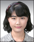
51세 여자 환자로 내원 1개월 전부터 우측 가슴이 심하게 부풀어 오르는 증상으로 내원하였다(Fig. 1). 환자는 내원 10년 전인 2002년에 유룬 주변 접근(periareolar approach)을 통한 큰가슴근하(subpectoral) 보형물 삽입으로 유방확대술을 시행 받았으며, 보형물에 대한 기록은 없었다. 양측 유방은 비대칭이었으며 좌측에 비해 우측이 부종이 심함 소견을 보였으나 압통이나 열감 등은 없었다.

Preoperative photograph of the patient. There was asymmetry, with right breast larger than the left.
보형물 파열이 의심되어 자기공명영상(MagneticResonance Imaging: MRI)검사를 시행하였다. 자기공명영상은 3T skyra (Simens, Germany)로 유방전용 코일을 사용하여 환자가 엎드린 자세로 시행하였으며 역동적 조영 증강 기법으로 시행하였다. 자기공명영상 검사상에서 우측 유방에 피막내 파열 및 피막외 파열이 있으며 피막외부 후면에 5x1.6 cm 의 육아종 이 있었다. 또한 액체-액체 층(fluid-fluid level)의 다른 신호 강도를 보이는 액체가 캡슐내에 고여 있어 다양한 단계의 출혈(hemorrhage) 이나 염증(inflammation) 등이 의심되었다 (Fig. 2). 환자는 양쪽 가슴의 보형물을 모두 제거하기를 원하였으며 추가적인 유방 확대술은 원하지 않았다. 전신마취하에 유방 주름하 접근법으로 피부 및 피하지방을 절개하자 피막(capsule) 내에 고여있던 검붉은 장액이 다량 나왔으며 우측 보형물은 파열되어 있었다. 좌측 보형물은 쭈글쭈글 하였으나(rumpled) 붕괴 없이 유지되고 있었다. 보형물은 오돌토돌한(textured) 형태였으며 자기 공명영상과 수술소견에서 하이드로겔 보형물이 의심되었다(Fig. 3). 피막도 완전히 제거하고 나서 늑골간 근육(intercostalis muscle) 과 피막 사이의 육아종성 종양을 제거하였다. 상단부 종양은 큰가슴근까지 연결되어 있었다(Fig. 4).병리조직 검사상에서 피막외 종양은 3.5×2.5×2.5cm의 육아종이었으며 피막은 육아조직과 섬유조직을 동반한 만성 염증 소견을 보였다. 환자는 수술 후 10개월까지 경과관찰하였으며 특별한 합병증은 관찰되지 않았다.

Images of the breast MRI. (Left) Axial view of a precontrast T1-weighted image. (Right) Axial view of a T1-weighted turbo spin echo (TSE) image. Both subpectoral implants are shown. An intracapsular rupture is shown, as is the large amount of fluid that collected around the ruptured implant. The fluid-fluid level demonstrates various stages of inflammation and hemorrhaging (arrows). The implant preserved its normal shape and contour without an inward folding of the shells, and it enlarged rather than collapsed. An extracapsular rupture was also identified, along with hydrogel extension to the pectoralis major, subcutaneous tissue, and intercostal area. There was a granuloma on the posterior portion of capsule (arrowheads).

Photograph of extracted implants. During Surgery, it was found that both implants were marked as PIP implants. The right implant was ruptured

Totally excised capsule and extracapsular mass. (Left) A capsule with an extracapsular mass was excised. The capsule’s internal surface shows a focal hyperemic appearance, which indicates chronic inflammation with granulation tissue and fibrosis. (Right) An extracapsular mass shows a fibrotic appearance correlated with pathologic results such as granulomatous inflammation.
III. 고 찰
유방성형술에서 보형물의 파열은 잘 알려진 합병증이다[8]. 최근 점차 보편적으로 사용되는 실리콘 보형물의 경우 세대를 거듭할수록 껍질의 강도와 내구성을 강화하여 실리콘 겔이 누출되거나 파열되지 않도록 개선되고 있다. 하지만 미국 식품의약국은 2006년 11월 유방성형을 목적으로 이용되는 코헤시브 겔 유방보형믈의 판매를 허가하면서 3년마다 자기공명영상을 촬영하도록 권유한 사실을 고려할 때 잠재적인 파열의 위험은 항상 염두에 두어야 한다. 자기공명영상은 실리콘 보형물 파열시 가장 특이도와 민감도가 높은 진단 방법으로 알려져 있어 실리콘 보형물 파열과 관련한 진단 및 치료에 대하여 많은 보고가 있어 왔다[9-14].
한편 하이드로겔 보형물은 식염수 보형물에 비해 점성이 높아 실리콘 사용이 금지되었던 2000년 전후에 우리나라에서 많이 사용되었다[1,2]. 이 보형물은 식염수 보형물에 비해 실리콘 외피가 얇고 약하게 제작되어 있는 경우가 있어서 파열의 빈도가 2배 이상 높은 것으로 알려져 있다[3].하이드로겔 보형물은 사용된 시기가 짧고 사용된 사람이 적어서 파열에 대한 보고가 적으며, 특히, 우리나라에서는 사용 실태에 대한 조사도 거의 없는 실정이다. 게다가 2000년 전 후에 수술을 받은 사람 중에는 증례의 환자처럼 본인이 하이드로겔 보형물을 사용하였다는 사실 자체를 인지하지 못하고 있는 경우도 있다.
Lee는[2] 부종과 크기 감소, 촉감변화 등으로 내원한 12명의 환자에서 수술을 통하여 피막내 파열 7명과 피막외 파열 5명의 케이스를 보고하였으며, Choi 등[3]은 양측 파열을 포함한 7 명의 환자에서 자기공명영상과 수술을 통하여 3명의 피막 내 파열과 4명의 피막외 파열을 보고하였다. 또한 영국, 한국, 네덜란드에서 절개부 분비물(discharge), 유방 크기 감소 및 증가 등의 증례가 보고된 적이 있었다.
Choi 등[3] 의 보고에 따르면 자기공명영상 상에서 피막내 파열의 경우 보형물 주변에 엑체집적(fluid collection)이 있으면서 보형물의 붕괴는 없으면서 보형물의 형태를 유지하고 있었으며 피막외 파열의 경우 보형물의 부분 혹은 전 붕괴를 동반하였으나 보형물 주변에 액체집적은 없이 오히려 피하 등에 액체집적이 있었다. 피막외 파열의 경우 하이드로겔은 큰 가슴근이나 늑간까지 퍼져있었다.
하이드로겔은 물에서 부풀어오르면서 수분을 함유하는 성질이 있어 주변 조직으로 쉽게 퍼져나간다[2,16]. 따라서, 누출시 수분을 흡수하게 되어 보형물 내부의 삼투압이 높아지면서 수분의 이동으로 보형물이 팽창하다가 외피가 마찰로 인한 스트레스를 견디다 못해 보형물 자체가 파열될 수 있으며[2-7], 수분의 함유 정도에 따라 자기공명영상에서도 다양한 신호강도(signal)로 보일 수 있다. 실제로 본원의 증례에서도 양측 유방내의 보형물 형태, 피막내 집적된 액체, 피막외 종괴의 신호강도가 모두 달랐다. 일반적으로 피막내 파열의 경우 하이드로겔이 누출되면서 생체내 수분을 흡수하게 되고 피막내로 퍼지면서 피막내에 액체 집적을 만들게 되며 피막 내부의 액체와 하이드로겔이 삼투압으로 이동하면서 양측이 등장성이 이루어지면 파열이 되더라도 붕괴되지 않는다. 피막외 파열의 경우는 피막내 파열 초기에 발견되지 못하고 방치되었다가 진행되는 것으로 알려져 왔다[3]. 따라서, 피막이 터지면서 파열 된 부위로 하이드로겔이 빠져나가면서 피막내에서 이루던 등장액을 이루지 못하고 보형물이 내부의 하이드로겔이 빠져나가면서 정도는 다르지만 붕괴되는 양상을 보인다. 본 증례는 자기 공명영상 상에서 피막내외 파열을 동시에 보였던 증례로 보형물의 형태를 유지면서 혈액과 염증이 섞여있는 두 층의 액체집적이 있었고 피막외에 하이드로겔이 나가서 만들어진 육아종성 종괴도 있었다. 또한 양측 보형물의 자기 공명영상의 신호강도가 다르고 신호강도가 다른 두 층의 액체집적이 있는 점은 하이드로겔의 삼투압설을 뒷받침하는 것으로 보인다, 피막외 종괴의 신호강도는 파열되지 않은 보형물내의 신호강도와 유사하여 하이드로겔의 누출에 의한 종괴임을 시사하였다.
피막내외 파열이 동시에 발견된 이유는 피막외 파열로 누출된 하이드로겔이 조기에 육아종을 이루면서 피막의 파열된 부위를 막았거나 피막내 파열에서 진행되는 조기에 발견되었기 때문으로 추정된다.
피막내 파열의 경우에는 식염수 세척만으로 제거가 가능하며 배액관을 유지하기도 한다. 피막외파열의 경우 주로 종물 형태로 누출되어 있으나 세포외액을 끌어들여 묽어진 형태로 존재하게 되므로 적은 양인 경우 세침천자(needle aspiration)을 시행할 수 있는 것으로 되어있으며[1], 육아종을 형성한 경우에는 이에 준하여 절제를 시행하여야 한다. 특별한 감염이 없는 경우 모든 문헌에서[1,2,3,7] 동시에 보형물을 재 삽입하여 문제가 없는 것으로 발표되었다. 그러나 제거만 시행한 경우에도 6개월 이상 부종이 지속되는 등[4] 다양한 임상적 소견을 보이는 만큼 재 삽입시 감염에 의한 구형구축 등을 예방하려면 신중한 판단이 우선되야 한다.
여러 논문에서 일반적으로 3~9년 사이에 파열을 감지하였으나 7년 이상 경과한 경우 조직 손상 범위가 크고 피막외 파열인 경우가 많았고 본 증례에서도 10년 경과된 환자였다[2-7].
2000년 12월 영국 MDA (Medical Devices Agency of the Department of Health)에 의해 하이드로겔 사용이 금지되었고 우리나라에서도 이후 사용이 중지되었지만 과거 이 보형물로 유방확대술을 받은 여성에 대한 조치가 필요할 것이다. 특히, PIP (Poly Implant Protsthesis, Seyne-sur-Mer, France)사의 하이드로겔 보형물인 경우 식약청의 승인 하에 정식으로 수입되어 유통된 바가 있어 이 보형물로 유방확대술을 받은 여성에 대한 교육과 검진이 필요할 것으로 보인다. 또한 본 증례의 환자처럼 보형물에 대한 정확한 인식이 없어 실리콘 보형물로 잘못 인지하고 있는 경우도 있으므로 하이드로겔 보형물 파열시의 증상이나 검사결과를 아는 것이 중요할 것이다.
IV. 결 론
하이드로겔 유방보형물은 쉽게 파열되는 성상이 있으며 파열 시 다양한 임상적 소견과 방사선학적 소견을 보이는 만큼 하이드로겔 유방보형물로 유방확대술을 시행한 경우 보형물 파손 여부에 대한 세심한 진찰이 필요하다.