


Effects of low-level laser therapy and adipose-derived stem cells on the viability of autologous fat grafts: a preliminary study
-
Chan Yeong Heo1
 , Young Soo Yoon2, Kyung Hee Min2, Sun Young Nam1, Kyu Sang Lee3, Byung Ho Shin1, Seunghee Lee1, Miji Lee1
- Received November 1, 2020 Revised January 28, 2021 Accepted March 19, 2021
- ABSTRACT
-
- Background
- Autologous fat grafts are commonly used in cosmetic and reconstructive surgery, and various methods are used to improve their viability. Low-level laser therapy (LLLT) can enhance the proliferation, growth, and differentiation of various cell lines, including stem cells. Our study investigated and compared the effects of LLLT and the addition of adipose-derived stem cells (ADSCs) on the viability of fat grafts.
- Methods
- Twenty nude mice were divided into four groups: control (group 1), LLLT irradiation (group 2), ADSC addition (group 3), and LLLT irradiation+ADSC addition (group 4). ADSCs were combined with the fat tissue. LLLT irradiation was performed once daily for 1 week from the day of grafting. After 8 weeks, the weight, volume, histology, and Western blot findings of the grafted fat tissues were evaluated.
- Results
- The retention rate and volume of the fat tissue in groups 2, 3, and 4 were higher than that of group 1, but the difference was not statistically significant. The number of capillaries, histological parameters, and immunofluorescence staining analyses for CD68, CD31, fibroblast growth factor, and vascular endothelial growth factor (VEGF) showed no significant differences among the four groups. The expression level of VEGF was higher in group 2 than in the other groups, but not to a statistically significant level.
- Conclusions
- LLLT and ADSCs did not significantly improve the viability of autologous fat grafts. Therefore, further study is necessary to develop safe and effective methods to improve the viability of these grafts for clinical application.
- INTRODUCTION
- INTRODUCTION
Low-level laser therapy (LLLT) is a type of phototherapy that uses low levels of red or near-infrared lasers with a wavelength of 600–1,100 nm and an output power of up to 500 mW. LLLT is athermally and atraumatically conducted through photoactivation of the target tissues or cells at low levels of photon energy [1,2]. LLLT has various biological effects, including prevention of cell apoptosis and stimulation of cell growth, proliferation, and differentiation [3-5]. Therefore, LLLT is clinically applied for wound healing, bone remodeling, nerve regeneration, pain attenuation, skin rejuvenation, and immune system modulation [1,6,7].Autologous fat grafts are ideal fillers in cosmetic and reconstructive surgery. Fat grafts have many advantages in that they are easy to harvest, abundant, cheap, and biocompatible [8-10]. Although clinical results have been improved through advances in fat grafting techniques, unpredictable absorption patterns remain challenging [8-12].Several efforts have been made to enhance the survival rate of grafted fat tissues, including the addition of growth factors and supplemental cells, such as adipose-derived stem cells (ADSCs). Cell-assisted lipotransfer (CAL), in which a stromal vascular fraction (SVF) or ADSCs are added to a fat graft, is widely performed in clinical settings [8,9,11-13]. The effects of LLLT on adipocytes and ADSCs have been investigated. LLLT enhances the viability and proliferation of ADSCs and stimulates vascular proliferation in adipose tissue [5,14]. Clinically, LLLT is used to enhance the viability of fat grafts [15].We therefore hypothesized that LLLT would enhance the survival rate of fat grafts and increase the effectiveness of ADSCs. Therefore, our study sought to investigate the effects of LLLT and ADSCs on the viability of grafted fat tissues using a nude mouse model.
- METHODS
- METHODS
- Harvesting of ADSCs
- Harvesting of ADSCs
Human fat tissue was resected from the remnant tissue of a transverse rectus abdominis musculocutaneous (TRAM) flap from a 56-year-old female patient (body mass index, 22.96 kg/m2). Approval for the use of human fat tissue was granted by the Institutional Review Board at Seoul National University Bundang Hospital. The fat tissues were washed, minced, and digested with 0.5% type I collagenase (Worthington, Lakewood, NJ, USA). The tissues were then washed with saline 5 times and centrifuged at 300 ×g for 5 minutes. Floating mature adipocytes were discarded. SVF was obtained from the precipitated pellets. ADSCs were seeded and cultured in Dulbecco’s modified Eagle’s medium containing 10% fetal bovine serum (Invitrogen-Gibco, Grand Island, NY, USA) supplemented with an antibiotic/antimycotic solution (Welgene Inc., Daegu, Korea) under normoxic conditions at 37°C. Cells from the third passage that were positive for the surface marker CD105 and negative for the surface markers CD19 and CD45 were used for experiments (Fig. 1).- Harvesting and preparing fat tissue
- Harvesting and preparing fat tissue
Human fat tissue was resected from the remnant tissue of a TRAM flap from a 49-year-old female patient (body mass index, 22.15 kg/m2). The tumescent solution was infiltrated into the subcutaneous layer of the resected fat tissue at a ratio of 2:1. The tumescent solution was composed of Ringer’s lactate solution (1,000 mL), 1:1,000 epinephrine (1 mL), 8.4% sodium bicarbonate (5 mL), and 1% lidocaine (50 mL).The fat tissue was then aspirated using a 14-gauge blunt cannula connected to the 10-mL syringe. The aspirated fat tissue was centrifuged at 1,800 ×g for 3 minutes. The aqueous bottom layer and the oily top layer were discarded. The prepared fat in the middle layer was transferred using a 1-mL syringe for fat grafting.- In vivo study
- In vivo study
Six-week-old male BALB/c-nude mice (n=20) were used in this experiment. The mice were divided into four groups, as shown in Table 1. In the control group (group 1), the fat tissue was grafted without ADSCs and irradiation was not performed. In group 2, the fat tissue was grafted without ADSCs and irradiation was performed. In group 3, fat tissues with ADSCs were grafted and the mice were not subjected to irradiation. In group 4, the fat tissue was grafted with ADSCs and the mice were subjected to irradiation. The fat tissue was grafted into the posterior nuchal area as a bolus, using an 18-gauge needle (Fig. 2A). The ADSCs (5×10⁶ cells/0.1 mL) were mixed with 0.9 mL of fat tissue. In groups 2 and 4, the mice were subjected to irradiation with an 830-nm Ga-Al-As laser (Healite II LED; Lutronic Co., Goyang, Korea) once a day for 1 week from the day of fat grafting (Fig. 2B). The power density of the laser was 40 mW/cm2. The energy density of the laser was 20 J/cm2.- Fat graft survival
- Fat graft survival
Fat graft survival was analyzed by assessing the weight and volume of the grafted fat tissue in the mice. The graft retention rate was calculated using the following equation; graft retention rate (%)=(postgraft weight/pre-graft weight)×100. The pre-graft weight (the net weight) of the grafted fat tissue was measured by subtracting the weight of an empty 1-mL syringe from that of a syringe containing the fat tissue for each fat graft.Eight weeks after the grafting was carried out, the mice were sacrificed, and the grafted fat tissue was harvested (Fig. 3). The harvested tissues were weighed and used to calculate the graft retention rate. To measure the volume of the harvested tissue, the liquid overflow method was used [16]. The tissue was placed in a 5-mL syringe containing 4 mL of saline, and the increase in the volume of the liquid was measured.- Histological analysis
- Histological analysis
The harvested fat tissues were fixed in paraformaldehyde solution for 2 days and then embedded in paraffin. Five-micrometer sections of the fat graft tissue were cut on a microtome (Microm, Walldorf, Germany) and stained with hematoxylin and eosin. Neovascularization was evaluated by counting the number of capillaries under ×40 magnification. The following histological parameters were analyzed semi-quantitatively: the presence of intact and nucleated fat cells, the presence of cysts and vacuoles, inflammation (infiltration of lymphocytes and macrophages), and the presence of fibrosis. Each parameter was graded on a scale of 0–5: 0 (absence), 1 (minimal presence), 2 (minimal to moderate presence), 3 (moderate presence), 4 (moderate to extensive presence), and 5 (extensive presence).The expression of macrophage infiltration (CD68), microvascular density (CD31), and the angiogenic factors fibroblast growth factor-2 (FGF-2) and vascular endothelial growth factor (VEGF) were measured in the harvested tissues using immunofluorescence staining. All antibodies were purchased from Abcam (Cambridge, UK).- Western blot analysis
- Western blot analysis
The expression of the angiogenic factor VEGF was measured in the harvested tissues using Western blotting.- Statistical analysis
- Statistical analysis
R language version 3.3.3 (R Foundation for Statistical Computing, Vienna, Austria) and T&F program version 3.0 (YooJin BioSoft, Goyang, Korea) were used for all statistical analyses. Data were expressed as median (first quartile to third quartile) for continuous variables, and as sample number and percentage for categorical variables. The Kruskal-Wallis H test was used to compare the survival rate, volume of transplanted fat tissue, and histologic parameters among the four treatment groups.
- RESULTS
- RESULTS
- Fat graft survival
- Fat graft survival
The graft retention rate of groups 2 (LLLT), 3 (ADSCs), and 4 (LLLT +ADSCs) was higher than that of group 1 (control). The retention rate of group 2 was higher than that of groups 3 and 4. However, the difference was not statistically significant (Fig. 4A). The volume of the fat grafts in groups 2, 3, and 4 was higher than that of group 1, but the difference was not statistically significant (Fig. 4B).- Histological analysis
- Histological analysis
The counting of capillaries showed no significant differences among the groups (Fig. 5A). The histological analysis of various parameters—namely, the presence of intact and nucleated fat cells, cysts and vacuoles, inflammation, and presence of fibrosis—showed no significant differences among the four groups (Table 2). Immunofluorescence staining analyses of CD68 (macrophage infiltration), CD31 (microvascular density), and angiogenic factors (FGF-2 and VEGF) also did not show any significant difference among the four groups (Fig. 5B).- Expression of angiogenic factors
- Expression of angiogenic factors
- DISCUSSION
- DISCUSSION
Various techniques have been used in attempts to overcome the unpredictable absorption patterns and increase the viability of autologous fat grafts. Of these, CAL has been the focus of a considerable amount of research. CAL enhances neo-vascularization and prevents fat graft complications, such as absorption, cyst formation, fibrosis, and necrosis [8,9,11-13]. The mechanism of CAL is not well understood. There are two main hypotheses with regard to its mode of action. One hypothesis is that ADSCs differentiate directly into adipocytes or endothelial cells. The other is that ADSCs secrete antiapoptotic and angiogenic growth factors, which improve fat graft volume retention and vascularization in the hypoxic graft environment. The latter hypothesis is currently accepted widely by researchers [11,17,18]. However, not all studies have reported positive effects of CAL. Peltoniemi et al. [19] reported that breast augmentation using CAL did not show any advantages in a comparative study. Conde-Green et al. [20] reported that CAL showed effects similar to those of other fat grafting techniques in an animal study. The study of ADSCs in fat grafts should take into account various factors including patient variability, the recipient site, the volume of fat aspiration, and the number of SVFs or ADSCs.CAL continues to have its limitations. The optimal dose of SVFs or ADSCs for CAL has not yet been determined. To obtain cells for CAL, patients have to sacrifice additional fat tissue. The safety of using SVF or ADSCs has yet to be evaluated. These cells are tumorigenic and can induce tumor growth through paracrine effects. Particularly in fat grafts with ADSCs or SVF for breast reconstruction following cancer, breast cancer recurrence should be taken into consideration. The proliferation of remaining cancer cells may be induced by ASDCs [21,22].LLLT induces biophysical effects on cells through the conversion of photon energy to chemical energy. LLLT can prevent cell apoptosis and stimulate cell growth, proliferation, and differentiation [2-5]. Numerous studies using various cell lines, including stem cells, and various parameters of LLLT have been reported [2,23]. Our previous study showed that LLLT enhanced the viability and proliferation of ADSCs both in vitro and in vivo [5]. Other studies have reported positive effects of LLLT on stem cells [24,25].In the present study, although statistically not significant, the LLLT-treated group showed a higher retention rate and VEGF expression than other groups. This result may reflect the photobioactivation caused by LLLT. It is believed that in fat grafts, LLLT increases cell proliferation and tissue oxygenation and modulates various cytokines and growth factors, both in adipocytes and in other cells, including ADSCs [14,15].LLLT enhances the viability and proliferation of ADSCs and stimulates vascular proliferation in adipose tissue [5,14]. Clinically, LLLT is used to enhance the viability of fat grafts [15].Owing to the use of low-energy lasers, LLLT does not emit heat or vibration [1]. Therefore, LLLT is unlikely to cause complications and can be used easily and safely in patients following fat grafting. However, to achieve optimal results with LLLT in fat grafts, further study is needed to determine the optimal parameters in various clinical applications.We did not find any significant difference in fat graft viability among the groups we studied, potentially because our sample size was small. Furthermore, since the skin thickness of nude mice is different from that of humans, the laser permeability may differ. Our study revealed that LLLT might not have an adequate effect on grafted fat tissue. Additionally, fat necrosis was found in the central portion of the grafted fat tissue. This may have influenced the weight of the grafted fat tissue, and a possible explanation is that the volume of fat graft might have been too large for the injection. Further experiments are required to address the limitations of this study.Herein, we were unable to conclusively identify a significant positive effect of LLLT and ADSCs on fat grafts in nude mice. Further investigations are needed to determine the safety and optimal method of CAL for clinical applications; meanwhile, LLLT may be a safer and easier method for fat grafts than CAL. However, to effectively apply LLLT to fat grafts, studies are needed to determine the most appropriate parameters.
- CONFLICTS OF INTEREST
- CONFLICTS OF INTEREST
No potential conflict of interest relevant to this article was reported.
- Notes
- Notes
-
Ethical approval The study was approved by the Institutional Review Board of the Seoul National University Bundang Hospital (IRB No. B-1608/358-302) and performed in accordance with the principles of the Declaration of Helsinki. The written informed consent was obtained. The animals were cared for, maintained, and treated in accordance with a protocol approved by the Institutional Animal Care and Use Committee of Seoul National University Bundang Hospital (IACUC No. BA1704-221/026-02).
Fig. 1.
Immunophenotyping of human adipose-derived stem cells. These cells were positive for the surface marker CD105, but negative for the hematopoietic cell-specific surface markers CD19 and CD45.
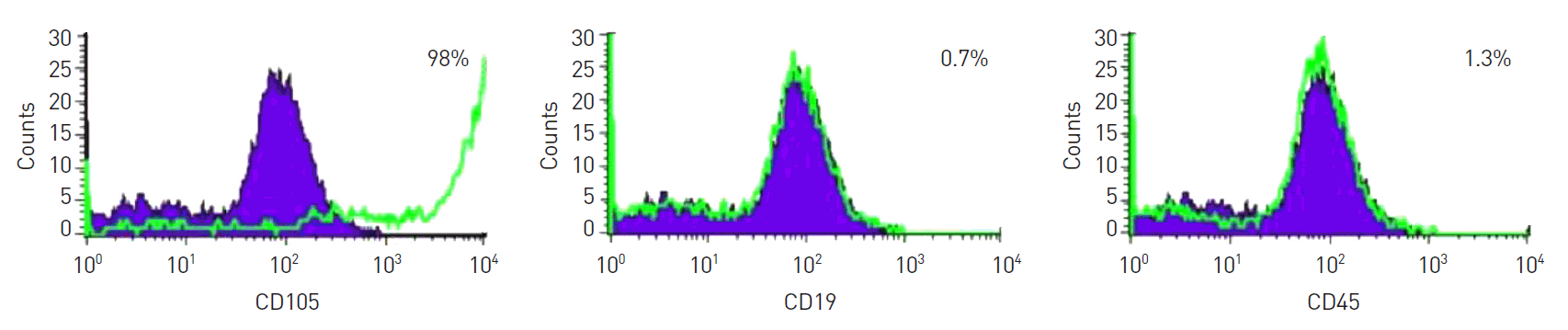
Fig. 2.
Animal experiment photos. (A) The fat tissue was grafted to the posterior nuchal area of the nude mouse. (B) Low-level laser therapy was applied using an 830 nm Ga-Al-As laser (B).

Fig. 3.
Animal experiment photos. (A) Eight weeks after fat grafting, the grafted fat was well maintained. (B) Grafted fat tissue was harvested.
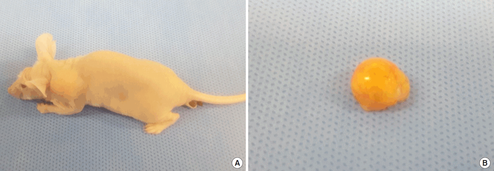
Fig. 4.
The graft retention rate (A), and volume of fat grafts for each group (B). First quartile, median, and third quartile are presented in the plot. LLLT, low-level laser therapy; ADSCs, adipose-derived stem cells.
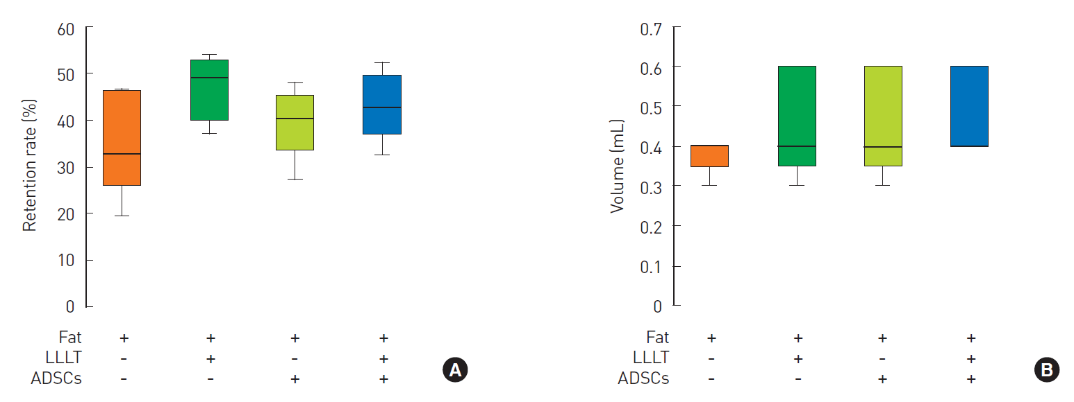
Fig. 5.
(A) Neovascularization was analyzed by counting the capillaries under ×40 magnification. (B) The expression of macrophage infiltration (CD68), microvascular density (CD31), and angiogenic factors (FGF-2 and VEGF) were measured using immunofluorescence staining. (C, D) The expression of the angiogenic factor VEGF was measured using Western blotting. LLLT, low-level laser therapy; ADSCs, adipose-derived stem cells; FGF-2, fibroblast growth factor-2; VEGF, vascular endothelial growth factor.

Table 1.
Experimental groups and protocols
Table 2.
Histological comparison of fat grafts among the four groups
- REFERENCES
- REFERENCES
- 1. Kim WS, Calderhead RG. Is light-emitting diode phototherapy (LED-LLLT) really effective? Laser Ther 2011;20:205–15.
[Article] [PubMed] [PMC]2. AlGhamdi KM, Kumar A, Moussa NA. Low-level laser therapy: a useful technique for enhancing the proliferation of various cultured cells. Lasers Med Sci 2012;27:237–49.
[Article] [PubMed]3. Conlan MJ, Rapley JW, Cobb CM. Biostimulation of wound healing by low-energy laser irradiation: a review. J Clin Periodontol 1996;23:492–6.
[Article] [PubMed]4. Huang YY, Chen AC, Carroll JD, et al. Biphasic dose response in low level light therapy. Dose Response 2009;7:358–83.
[Article] [PubMed] [PMC]5. Min KH, Byun JH, Heo CY, et al. Effect of low-level laser therapy on human adipose-derived stem cells: in vitro and in vivo studies. Aesthetic Plast Surg 2015;39:778–82.
[Article] [PubMed]6. Calderhead RG, Kubota J, Trelles MA, et al. One mechanism behind LED phototherapy for wound healing and skin rejuvenation: key role of the mast cell. Laser Ther 2008;17:141–8.
[Article]7. Goldberg DJ, Amin S, Russell BA, et al. Combined 633-nm and 830-nm led treatment of photoaging skin. J Drugs Dermatol 2006;5:748–53.
[PubMed]8. Paik KJ, Zielins ER, Atashroo DA, et al. Studies in fat grafting: part V. Cell-assisted lipotransfer to enhance fat graft retention is dose dependent. Plast Reconstr Surg 2015;136:67–75.9. Lu F, Li J, Gao J, et al. Improvement of the survival of human autologous fat transplantation by using VEGF-transfected adipose-derived stem cells. Plast Reconstr Surg 2009;124:1437–46.
[Article] [PubMed]10. Minn KW, Min KH, Chang H, et al. Effects of fat preparation methods on the viabilities of autologous fat grafts. Aesthetic Plast Surg 2010;34:626–31.
[Article] [PubMed]11. Toyserkani NM, Quaade ML, Sorensen JA. Cell-assisted lipotransfer: a systematic review of its efficacy. Aesthetic Plast Surg 2016;40:309–18.
[Article] [PubMed] [PMC]12. Zielins ER, Brett EA, Blackshear CP, et al. Purified adipose-derived stromal cells provide superior fat graft retention compared with unenriched stromal vascular fraction. Plast Reconstr Surg 2017;139:911–4.
[Article] [PubMed] [PMC]13. Hamed S, Egozi D, Kruchevsky D, et al. Erythropoietin improves the survival of fat tissue after its transplantation in nude mice. PLoS One 2010;5:e13986.
[Article] [PubMed] [PMC]14. Brown SA, Rohrich RJ, Kenkel J, et al. Effect of low-level laser therapy on abdominal adipocytes before lipoplasty procedures. Plast Reconstr Surg 2004;113:1796–804.
[Article] [PubMed]15. Nita AC, Jianu DM, Florescu IP, et al. The synergy between lasers and adipose tissues surgery in cervicofacial rejuvenation: histopathological aspects. Rom J Morphol Embryol 2013;54:1039–43.
[PubMed]16. Ayhan M, Senen D, Adanali G, et al. Use of beta blockers for increasing survival of free fat grafts. Aesthetic Plast Surg 2001;25:338–42.
[Article] [PubMed]17. Rinker BD, Vyas KS. Do stem cells have an effect when we fat graft? Ann Plast Surg 2016;76 Suppl 4:S359–63.
[Article] [PubMed]18. Garza RM, Rennert RC, Paik KJ, et al. Studies in fat grafting: part IV. Adipose-derived stromal cell gene expression in cell-assisted lipotransfer. Plast Reconstr Surg 2015;135:1045–55.19. Peltoniemi HH, Salmi A, Miettinen S, et al. Stem cell enrichment does not warrant a higher graft survival in lipofilling of the breast: a prospective comparative study. J Plast Reconstr Aesthet Surg 2013;66:1494–503.
[Article] [PubMed]20. Conde-Green A, Wu I, Graham I, et al. Comparison of 3 techniques of fat grafting and cell-supplemented lipotransfer in athymic rats: a pilot study. Aesthet Surg J 2013;33:713–21.
[PubMed]21. Rowan BG, Gimble JM, Sheng M, et al. Human adipose tissue-derived stromal/stem cells promote migration and early metastasis of triple negative breast cancer xenografts. PLoS One 2014;9:e89595.
[Article] [PubMed] [PMC]22. Bertolini F, Petit JY, Kolonin MG. Stem cells from adipose tissue and breast cancer: hype, risks and hope. Br J Cancer 2015;112:419–23.
[Article] [PubMed] [PMC]23. Mussttaf RA, Jenkins DFL, Jha AN. Assessing the impact of low level laser therapy (LLLT) on biological systems: a review. Int J Radiat Biol 2019;95:120–43.
[Article] [PubMed]